


 癌症的基本特征包括细胞增殖、血管生成、迁移、凋亡逃避机制和细胞永生等。找到癌症发生过程中这些通路的关键标记物和对应的抗体用于检测至关重要。
 为您推荐一个泛素化位点预测神器——泛素化分析工具,可以为您的蛋白的泛素化位点作出预测和评分。
 细胞自噬受体图形绘图工具为你的蛋白的细胞受体结合位点作出预测和评分,识别结合到自噬通路中的蛋白是非常重要的,便于让我们理解自噬在正常生理、病理过程中的作用,如发育、细胞分化、神经退化性疾病、压力条件下、感染和癌症。
CEA(C3) Mouse mAb
CEA(C3) Mouse mAb
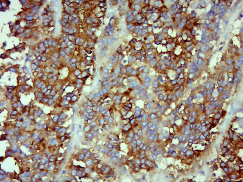
- 产品详情
- 实验流程
- 背景知识
Application 
| IHC-P, IHC-F, IF |
|---|---|
| Primary Accession | P06731 |
| Reactivity | Human |
| Host | Mouse |
| Clonality | Monoclonal |
| Calculated MW | 76796 Da |
| Physical State | Liquid |
| Immunogen | KLH conjugated synthetic peptide derived from human CEA/CD66e/CEACAM5 |
| Epitope Specificity | full length |
| Isotype | IgG |
| Purity | affinity purified by Protein A |
| Buffer | 0.01M PBS (pH7.4) with 0.02% Proclin300. |
| SUBCELLULAR LOCATION | Cell membrane; Lipid-anchor, GPI-anchor |
| Important Note | This product as supplied is intended for research use only, not for use in human, therapeutic or diagnostic applications. |
| Background Descriptions | CEA-related cell adhesion molecules (CEACAM) belong to the carcinoembryonic antigen (CEA) family. It consists of seven CEACAM (CEACAM 1, CEACAM 3-CEACAM 8) and 11 pregnancy-specific glyco-protein (PSG 1-PSG 11) members. The CEA family proteins belong to the immunoglobulin (Ig) superfamily and are composed of one Ig variable-like (IgV) and a varying number (0-6) of Ig constant-like (IgC) domains. CEACAM molecules are membrane-bound either via a transmembrane domain or a glycosyl phosphatidyl inositol (GPI) anchor. CEACAM molecules are differentially expressed in epithelial cells or in leucocytes. Over-expression of CEA/ CEACAM 5 in tumors of epithelial origin is the basis of its wide-spread use as a tumor marker. The function of CEACAM family members varies widely: they function as cell adhesion molecules, tumor suppressors, regulators of lymphocyte and dendritic cell activation, receptors of Neisseria species and other bacteria.CEA (Carcino Embryonic Antigen, CD66e) is synthesized during development in the fetal gut, and re-expressed in increased amounts in intestinal carcinomas and several other tumors. CEA is a member of carcinoembryonic antigens, immunoglobulin supergene family and consists of a single N domain (structural homology to the immunoglobulin variable) and six immunoglobulin constant-like A (A1, A2, A3) and B domains (B1, B2, B3). Antibodies to CEA are useful in identifying the origin of various metastatic adenocarcinomas and in distinguishing pulmonary adenocarcinomas (60 to 70% are CEA+) from pleural mesotheliomas (rarely or weakly CEA+). CEA is a member of a large family of glycoproteins, a useful tumor marker for adenocarcinoma, and found in adenocarcinomas of endodermally derived digestive system epithelium and fetal colon. Two subgroups of the CEA family, the CEA cell adhesion molecules and the pregnancy-specific glycoproteins, are located within a 1.2 Mb cluster on the long arm of chromosome 19. Eleven pseudogenes of the CEA cell adhesion molecule subgroup are also found in the cluster. CEA was originally described in bile ducts of liver as biliary glycoprotein. Subsequently, CEA was found to be a cell-cell adhesion molecule detected on leukocytes, epithelia, and endothelia. The encoded protein mediates cell adhesion via homophilic as well as heterophilic binding to other proteins of the subgroup. Multiple cellular activities have been attributed to the encoded protein, including roles in the differentiation and arrangement of tissue three-dimensional structure, angiogenesis, apoptosis, tumor suppression, metastasis, and the modulation of innate and adaptive immune responses. Multiple transcript variants encoding different isoforms have been reported, but the full-length nature of all variants has not been defined. |
| Gene ID | 1048 |
|---|---|
| Other Names | Cell adhesion molecule CEACAM5, Carcinoembryonic antigen, CEA, Carcinoembryonic antigen-related cell adhesion molecule 5, CEA cell adhesion molecule 5 {ECO:0000312|HGNC:HGNC:1817}, Meconium antigen 100, CD66e, CEACAM5 (HGNC:1817) |
| Dilution | IHC-P=1:100-500,IHC-F=1:100-50javascript:;0,IF=1:100-500,ELISA=1:5000-10000 |
| Storage | Store at -20 °C for one year. Avoid repeated freeze/thaw cycles. When reconstituted in sterile pH 7.4 0.01M PBS or diluent of antibody the antibody is stable for at least two weeks at 2-4 °C. |
For Research Use Only. Not For Use In Diagnostic Procedures.
| Name | CEACAM5 (HGNC:1817) |
|---|---|
| Function | Cell surface glycoprotein that plays a role in cell adhesion, intracellular signaling and tumor progression (PubMed:10864933, PubMed:10910050, PubMed:2803308). Mediates homophilic and heterophilic cell adhesion with other carcinoembryonic antigen-related cell adhesion molecules, such as CEACAM6 (PubMed:2803308). Plays a role as an oncogene by promoting tumor progression; induces resistance to anoikis of colorectal carcinoma cells (PubMed:10910050). |
| Cellular Location | Cell membrane; Lipid-anchor, GPI-anchor; Extracellular side. Apical cell membrane. Cell surface Note=Localized to the apical glycocalyx surface |
| Tissue Location | Expressed in columnar epithelial and goblet cells of the colon (at protein level) (PubMed:10436421). Found in adenocarcinomas of endodermally derived digestive system epithelium and fetal colon. |
Provided below are standard protocols that you may find useful for product applications.
BACKGROUND
This product as supplied is intended for research use only, not for use in human, therapeutic or diagnostic applications.
REFERENCES
Kuroki M.,et al.J. Biol. Chem. 266:11810-11817(1991).
Nagel G.,et al.Eur. J. Biochem. 214:27-35(1993).
Grimwood J.,et al.Nature 428:529-535(2004).
Mural R.J.,et al.Submitted (SEP-2005) to the EMBL/GenBank/DDBJ databases.
Khan W.N.,et al.Genomics 14:384-390(1992).

终于等到您。ABCEPTA(百远生物)抗体产品。
点击下方“我要评价 ”按钮提交您的反馈信息,您的反馈和评价是我们最宝贵的财富之一,
我们将在1-3个工作日内处理您的反馈信息。
如有疑问,联系:0512-88856768 tech-china@abcepta.com.


