


 癌症的基本特征包括细胞增殖、血管生成、迁移、凋亡逃避机制和细胞永生等。找到癌症发生过程中这些通路的关键标记物和对应的抗体用于检测至关重要。
 为您推荐一个泛素化位点预测神器——泛素化分析工具,可以为您的蛋白的泛素化位点作出预测和评分。
 细胞自噬受体图形绘图工具为你的蛋白的细胞受体结合位点作出预测和评分,识别结合到自噬通路中的蛋白是非常重要的,便于让我们理解自噬在正常生理、病理过程中的作用,如发育、细胞分化、神经退化性疾病、压力条件下、感染和癌症。
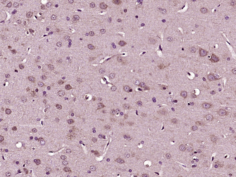
RhoA Rabbit pAb
RhoA Rabbit pAb
- 产品详情
- 实验流程
- 背景知识
Application
| IHC-P, IHC-F, IF |
|---|---|
| Primary Accession | P61586 |
| Reactivity | Human, Mouse, Rat |
| Predicted | Chicken, Dog, Rabbit, Zebrafish |
| Host | Rabbit |
| Clonality | Polyclonal |
| Calculated MW | 21768 Da |
| Physical State | Liquid |
| Immunogen | KLH conjugated synthetic peptide derived from human RhoA |
| Epitope Specificity | 101-193/193 |
| Isotype | IgG |
| Purity | affinity purified by Protein A |
| Buffer | 0.01M TBS (pH7.4) with 1% BSA, 0.02% Proclin300 and 50% Glycerol. |
| SUBCELLULAR LOCATION | Cell membrane; Lipid-anchor; Cytoplasmic side. Cytoplasm, cytoskeleton. Cleavage furrow. Cytoplasm, cell cortex. Midbody. Note=Localized to cell-cell contacts in calcium-treated keratinocytes. Translocates to the equatorial region before furrow formation in a ECT2-dependent manner. Localizes to the equatorial cell cortex (at the site of the presumptive furrow) in early anaphase in a activated form and in a myosin- and actin-independent manner. |
| SIMILARITY | Belongs to the small GTPase superfamily. Rho family. |
| SUBUNIT | Interacts with ARHGEF28. Binds PRKCL1, ROCK1 and ROCK2. Interacts with ARHGEF2, ARHGEF3, NET1 and RTKN. Interacts with PLCE1 and AKAP13. Interacts (in the constitutively activated, GTP-bound form) with DGKQ. Interacts with human respiratory syncytial virus (HRSV) protein F; this interaction facilitates virus-induced syncytium formation. Interacts with GNB2L1/RACK1; enhances RHOA activation. Interacts with PKP4; the interaction is detected at the midbody. Interacts (GTP-bound form preferentially) with PKN2; the interaction stimulates autophosphorylation and phosphorylation of PKN2. |
| Post-translational modifications | Substrate for botulinum ADP-ribosyltransferase. Cleaved by yopT protease when the cell is infected by some Yersinia pathogens. This removes the lipid attachment, and leads to its displacement from plasma membrane and to subsequent cytoskeleton cleavage. AMPylation at Tyr-34 and Thr-37 are mediated by bacterial enzymes in case of infection by H.somnus and V.parahaemolyticus, respectively. AMPylation occurs in the effector region and leads to inactivation of the GTPase activity by preventing the interaction with downstream effectors, thereby inhibiting actin assembly in infected cells. It is unclear whether some human enzyme mediates AMPylation; FICD has such ability in vitro but additional experiments remain to be done to confirm results in vivo. Phosphorylation by PRKG1 at Ser-188 inactivates RHOA signaling. Ubiquitinated by the BCR(BACURD1) and BCR(BACURD2) E3 ubiquitin ligase complexes, leading to its degradation by the proteasome, thereby regulating the actin cytoskeleton and cell migration. |
| Important Note | This product as supplied is intended for research use only, not for use in human, therapeutic or diagnostic applications. |
| Background Descriptions | This gene encodes a member of the Rho family of small GTPases, which cycle between inactive GDP-bound and active GTP-bound states and function as molecular switches in signal transduction cascades. Rho proteins promote reorganization of the actin cytoskeleton and regulate cell shape, attachment, and motility. The protein encoded by this gene is prenylated at its C-terminus, and localizes to the cytoplasm and plasma membrane. It is thought to be important in cell locomotion. Overexpression of this gene is associated with tumor cell proliferation and metastasis. Multiple alternatively spliced variants, encoding the same protein, have been identified. |
| Gene ID | 387 |
|---|---|
| Other Names | Transforming protein RhoA, 3.6.5.2, Rho cDNA clone 12, h12, RHOA (HGNC:667), ARH12, ARHA, RHO12 |
| Target/Specificity | Expressed highly in placenta, prostate and trachea and lower expression in the small intestine and lung. |
| Dilution | IHC-P=1:100-500,IHC-F=1:100-500,IF=1:200-800 |
| Storage | Store at -20 °C for one year. Avoid repeated freeze/thaw cycles. When reconstituted in sterile pH 7.4 0.01M PBS or diluent of antibody the antibody is stable for at least two weeks at 2-4 °C. |
| Name | RHOA (HGNC:667) |
|---|---|
| Synonyms | ARH12, ARHA, RHO12 |
| Function | Small GTPase which cycles between an active GTP-bound and an inactive GDP-bound state. Mainly associated with cytoskeleton organization, in active state binds to a variety of effector proteins to regulate cellular responses such as cytoskeletal dynamics, cell migration and cell cycle (PubMed:23871831). Regulates a signal transduction pathway linking plasma membrane receptors to the assembly of focal adhesions and actin stress fibers (PubMed:31570889, PubMed:8910519, PubMed:9121475). Involved in a microtubule-dependent signal that is required for the myosin contractile ring formation during cell cycle cytokinesis (PubMed:12900402, PubMed:16236794). Plays an essential role in cleavage furrow formation. Required for the apical junction formation of keratinocyte cell-cell adhesion (PubMed:20974804, PubMed:23940119). Essential for the SPATA13-mediated regulation of cell migration and adhesion assembly and disassembly (PubMed:19934221). The MEMO1-RHOA-DIAPH1 signaling pathway plays an important role in ERBB2- dependent stabilization of microtubules at the cell cortex. It controls the localization of APC and CLASP2 to the cell membrane, via the regulation of GSK3B activity. In turn, membrane-bound APC allows the localization of the MACF1 to the cell membrane, which is required for microtubule capture and stabilization (PubMed:20937854). Involved in the reorientation of endothelial cells and their actin stress fibers in response to cellular mechantransduction-mediated activation by ARHGEF40 (By similarity). Regulates KCNA2 potassium channel activity by reducing its location at the cell surface in response to CHRM1 activation; promotes KCNA2 endocytosis (PubMed:19403695, PubMed:9635436). Acts as an allosteric activator of guanine nucleotide exchange factor ECT2 by binding in its activated GTP-bound form to the PH domain of ECT2 which stimulates the release of PH inhibition and promotes the binding of substrate RHOA to the ECT2 catalytic center (PubMed:31888991). May be an activator of PLCE1 (PubMed:16103226). In neurons, involved in the inhibition of the initial spine growth. Upon activation by CaMKII, modulates dendritic spine structural plasticity by relaying CaMKII transient activation to synapse-specific, long-term signaling (By similarity). Acts as a regulator of platelet alpha-granule release during activation and aggregation of platelets (By similarity). When activated by DAAM1 may signal centrosome maturation and chromosomal segregation during cell division. May also be involved in contractile ring formation during cytokinesis. |
| Cellular Location | Cell membrane; Lipid-anchor; Cytoplasmic side. Cytoplasm, cytoskeleton. Cleavage furrow. Cytoplasm, cell cortex. Midbody. Cell projection, lamellipodium {ECO:0000250|UniProtKB:Q9QUI0}. Cell projection, dendrite {ECO:0000250|UniProtKB:Q9QUI0}. Nucleus Cytoplasm. Note=Localized to cell-cell contacts in calcium-treated keratinocytes (By similarity). Translocates to the equatorial region before furrow formation in a ECT2-dependent manner. Localizes to the equatorial cell cortex (at the site of the presumptive furrow) in early anaphase in an activated form and in a myosin- and actin-independent manner. Colocalizes with KANK1 at the contractile ring. Colocalizes with DAAM1 and KANK1 around centrosomes {ECO:0000250|UniProtKB:Q9QUI0} |
For Research Use Only. Not For Use In Diagnostic Procedures.
Provided below are standard protocols that you may find useful for product applications.
BACKGROUND
This gene encodes a member of the Rho family of small GTPases, which cycle between inactive GDP-bound and active GTP-bound states and function as molecular switches in signal transduction cascades. Rho proteins promote reorganization of the actin cytoskeleton and regulate cell shape, attachment, and motility. The protein encoded by this gene is prenylated at its C-terminus, and localizes to the cytoplasm and plasma membrane. It is thought to be important in cell locomotion. Overexpression of this gene is associated with tumor cell proliferation and metastasis. Multiple alternatively spliced variants, encoding the same protein, have been identified.
REFERENCES
Yeramian P.,et al.Nucleic Acids Res. 15:1869-1869(1987).
Fagan K.P.,et al.Exp. Eye Res. 59:235-237(1994).
Puhl H.L. III,et al.Submitted (APR-2002) to the EMBL/GenBank/DDBJ databases.
Kalnine N.,et al.Submitted (OCT-2004) to the EMBL/GenBank/DDBJ databases.
Suzuki Y.,et al.Submitted (APR-2005) to the EMBL/GenBank/DDBJ databases.
终于等到您。ABCEPTA(百远生物)抗体产品。
点击下方“我要评价 ”按钮提交您的反馈信息,您的反馈和评价是我们最宝贵的财富之一,
我们将在1-3个工作日内处理您的反馈信息。
如有疑问,联系:0512-88856768 tech-china@abcepta.com.


