


 癌症的基本特征包括细胞增殖、血管生成、迁移、凋亡逃避机制和细胞永生等。找到癌症发生过程中这些通路的关键标记物和对应的抗体用于检测至关重要。
 为您推荐一个泛素化位点预测神器——泛素化分析工具,可以为您的蛋白的泛素化位点作出预测和评分。
 细胞自噬受体图形绘图工具为你的蛋白的细胞受体结合位点作出预测和评分,识别结合到自噬通路中的蛋白是非常重要的,便于让我们理解自噬在正常生理、病理过程中的作用,如发育、细胞分化、神经退化性疾病、压力条件下、感染和癌症。
ERK1/2 Rabbit pAb
ERK1/2 Rabbit pAb

- 产品详情
- 实验流程
- 背景知识
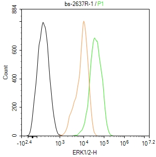
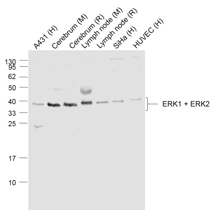
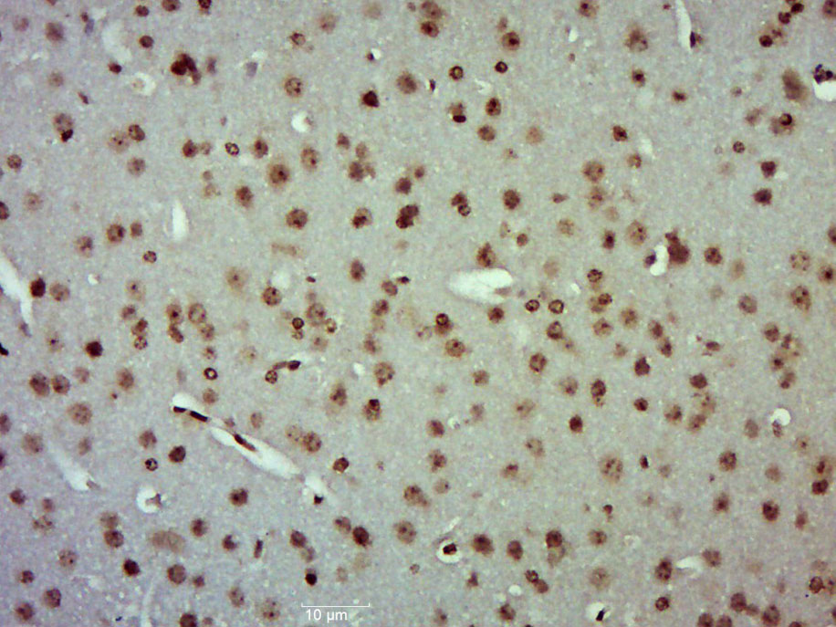
Application
| WB, IHC-P, IHC-F, IF |
|---|---|
| Primary Accession | P27361 |
| Reactivity | Human, Mouse, Rat |
| Predicted | Chicken, Dog, Pig, Horse, Rabbit, Sheep, Goat |
| Host | Rabbit |
| Clonality | Polyclonal |
| Calculated MW | 43136 Da |
| Physical State | Liquid |
| Immunogen | KLH conjugated synthetic peptide derived from human ERK1/2 |
| Epitope Specificity | 251-358/358 |
| Isotype | IgG |
| Purity | affinity purified by Protein A |
| Buffer | 0.01M TBS (pH7.4) with 1% BSA, 0.02% Proclin300 and 50% Glycerol. |
| SUBCELLULAR LOCATION | Cytoplasm, cytoskeleton, spindle. Nucleus. Cytoplasm, cytoskeleton, centrosome. Cytoplasm. Note=Associated with the spindle duringprometaphase and metaphase. PEA15-binding andphosphorylated DAPK1 promote its cytoplasmic retention.Phosphorylation at Ser-244 and Ser-246 as well asautophosphorylation at Thr-188 promote nuclear localization. |
| SIMILARITY | Belongs to the protein kinase superfamily. CMGCSer/Thr protein kinase family. MAP kinase subfamily. Contains 1 protein kinase domain. |
| SUBUNIT | Binds both upstream activators and downstream substratesin multimolecular complexes. Interacts with ADAM15, ARHGEF2, ARRB2,DAPK1 (via death domain), HSF4, IER3, IPO7, DUSP6, NISCH, SGK1, andisoform 1 of NEK2. Interacts (via phosphorylated form) with TPR(via C-terminus region and phosphorylated form); the interactionrequires dimerization of MAPK1/ERK2 and increases following EGFstimulation. Interacts (phosphorylated form) withCAV2 ('Tyr-19'-phosphorylated form); the interaction, promoted byinsulin, leads to nuclear location and MAPK1 activation. Interacts with DCC. Interacts withMORG1, PEA15 and MKNK2. MKNK2 isoform 1 binding prevents fromdephosphorylation and inactivation. The phosphorylated forminteracts with PML. |
| Post-translational modifications | Dually phosphorylated on Thr-183 and Tyr-185, which activatesthe enzyme. Ligand-activated ALK induces tyrosine phosphorylation. Dephosphorylated by PTPRJ at Tyr-185. Phosphorylated upon FLT3 and KIT signaling. |
| Important Note | This product as supplied is intended for research use only, not for use in human, therapeutic or diagnostic applications. |
| Background Descriptions | The protein encoded by this gene is a member of the MAPkinase family. MAP kinases, also known as extracellularsignal-regulated kinases (ERKs), act in a signaling cascade thatregulates various cellular processes such as proliferation,differentiation, and cell cycle progression in response to avariety of extracellular signals. This kinase is activated byupstream kinases, resulting in its translocation to the nucleuswhere it phosphorylates nuclear targets. Alternatively splicedtranscript variants encoding different protein isoforms have beendescribed. [provided by RefSeq, Jul 2008]. |
| Gene ID | 5595 |
|---|---|
| Other Names | Mitogen-activated protein kinase 3, MAP kinase 3, MAPK 3, 2.7.11.24, ERT2, Extracellular signal-regulated kinase 1, ERK-1, Insulin-stimulated MAP2 kinase, MAP kinase isoform p44, p44-MAPK, Microtubule-associated protein 2 kinase, p44-ERK1, MAPK3, ERK1, PRKM3 |
| Target/Specificity | Widely expressed. |
| Dilution | WB=1:1000-5000,IHC-P=1:100-500,IHC-F=1:100-500,ICC/IF=1:50-200,IF=1:100-500,Flow-Cyt=1 µg/Test |
| Storage | Store at -20 °C for one year. Avoid repeated freeze/thaw cycles. When reconstituted in sterile pH 7.4 0.01M PBS or diluent of antibody the antibody is stable for at least two weeks at 2-4 °C. |
| Name | MAPK3 |
|---|---|
| Synonyms | ERK1, PRKM3 |
| Function | Serine/threonine kinase which acts as an essential component of the MAP kinase signal transduction pathway (PubMed:34497368). MAPK1/ERK2 and MAPK3/ERK1 are the 2 MAPKs which play an important role in the MAPK/ERK cascade. They participate also in a signaling cascade initiated by activated KIT and KITLG/SCF. Depending on the cellular context, the MAPK/ERK cascade mediates diverse biological functions such as cell growth, adhesion, survival and differentiation through the regulation of transcription, translation, cytoskeletal rearrangements. The MAPK/ERK cascade also plays a role in initiation and regulation of meiosis, mitosis, and postmitotic functions in differentiated cells by phosphorylating a number of transcription factors. About 160 substrates have already been discovered for ERKs. Many of these substrates are localized in the nucleus, and seem to participate in the regulation of transcription upon stimulation. However, other substrates are found in the cytosol as well as in other cellular organelles, and those are responsible for processes such as translation, mitosis and apoptosis. Moreover, the MAPK/ERK cascade is also involved in the regulation of the endosomal dynamics, including lysosome processing and endosome cycling through the perinuclear recycling compartment (PNRC); as well as in the fragmentation of the Golgi apparatus during mitosis. The substrates include transcription factors (such as ATF2, BCL6, ELK1, ERF, FOS, HSF4 or SPZ1), cytoskeletal elements (such as CANX, CTTN, GJA1, MAP2, MAPT, PXN, SORBS3 or STMN1), regulators of apoptosis (such as BAD, BTG2, CASP9, DAPK1, IER3, MCL1 or PPARG), regulators of translation (such as EIF4EBP1) and a variety of other signaling-related molecules (like ARHGEF2, DEPTOR, FRS2 or GRB10) (PubMed:35216969). Protein kinases (such as RAF1, RPS6KA1/RSK1, RPS6KA3/RSK2, RPS6KA2/RSK3, RPS6KA6/RSK4, SYK, MKNK1/MNK1, MKNK2/MNK2, RPS6KA5/MSK1, RPS6KA4/MSK2, MAPKAPK3 or MAPKAPK5) and phosphatases (such as DUSP1, DUSP4, DUSP6 or DUSP16) are other substrates which enable the propagation the MAPK/ERK signal to additional cytosolic and nuclear targets, thereby extending the specificity of the cascade. Phosphorylates GJA1 at 'Ser-279' and 'Ser-282' resulting in an increase in GJA1 ubiquitination and ultimately lysosomal degradation (By similarity). |
| Cellular Location | Cytoplasm {ECO:0000250|UniProtKB:P21708}. Nucleus. Membrane, caveola {ECO:0000250|UniProtKB:P21708}. Cell junction, focal adhesion {ECO:0000250|UniProtKB:Q63844} Note=Autophosphorylation at Thr-207 promotes nuclear localization (PubMed:19060905). PEA15-binding redirects the biological outcome of MAPK3 kinase-signaling by sequestering MAPK3 into the cytoplasm (By similarity). {ECO:0000250|UniProtKB:Q63844, ECO:0000269|PubMed:19060905} |
For Research Use Only. Not For Use In Diagnostic Procedures.
Provided below are standard protocols that you may find useful for product applications.
BACKGROUND
The protein encoded by this gene is a member of the MAPkinase family. MAP kinases, also known as extracellularsignal-regulated kinases (ERKs), act in a signaling cascade thatregulates various cellular processes such as proliferation,differentiation, and cell cycle progression in response to avariety of extracellular signals. This kinase is activated byupstream kinases, resulting in its translocation to the nucleuswhere it phosphorylates nuclear targets. Alternatively splicedtranscript variants encoding different protein isoforms have beendescribed. [provided by RefSeq, Jul 2008].
REFERENCES
Her J.-H.,et al.Nucleic Acids Res. 19:3743-3743(1991).
Carninci P.,et al.Science 309:1559-1563(2005).
Lubec G.,et al.Submitted (JAN-2009) to UniProtKB.
Ershler M.A.,et al.Gene 124:305-306(1993).
Payne D.M.,et al.EMBO J. 10:885-892(1991).
终于等到您。ABCEPTA(百远生物)抗体产品。
点击下方“我要评价 ”按钮提交您的反馈信息,您的反馈和评价是我们最宝贵的财富之一,
我们将在1-3个工作日内处理您的反馈信息。
如有疑问,联系:0512-88856768 tech-china@abcepta.com.


