


 癌症的基本特征包括细胞增殖、血管生成、迁移、凋亡逃避机制和细胞永生等。找到癌症发生过程中这些通路的关键标记物和对应的抗体用于检测至关重要。
 为您推荐一个泛素化位点预测神器——泛素化分析工具,可以为您的蛋白的泛素化位点作出预测和评分。
 细胞自噬受体图形绘图工具为你的蛋白的细胞受体结合位点作出预测和评分,识别结合到自噬通路中的蛋白是非常重要的,便于让我们理解自噬在正常生理、病理过程中的作用,如发育、细胞分化、神经退化性疾病、压力条件下、感染和癌症。
Lipocalin 2/NGAL Rabbit pAb
Lipocalin 2/NGAL Rabbit pAb
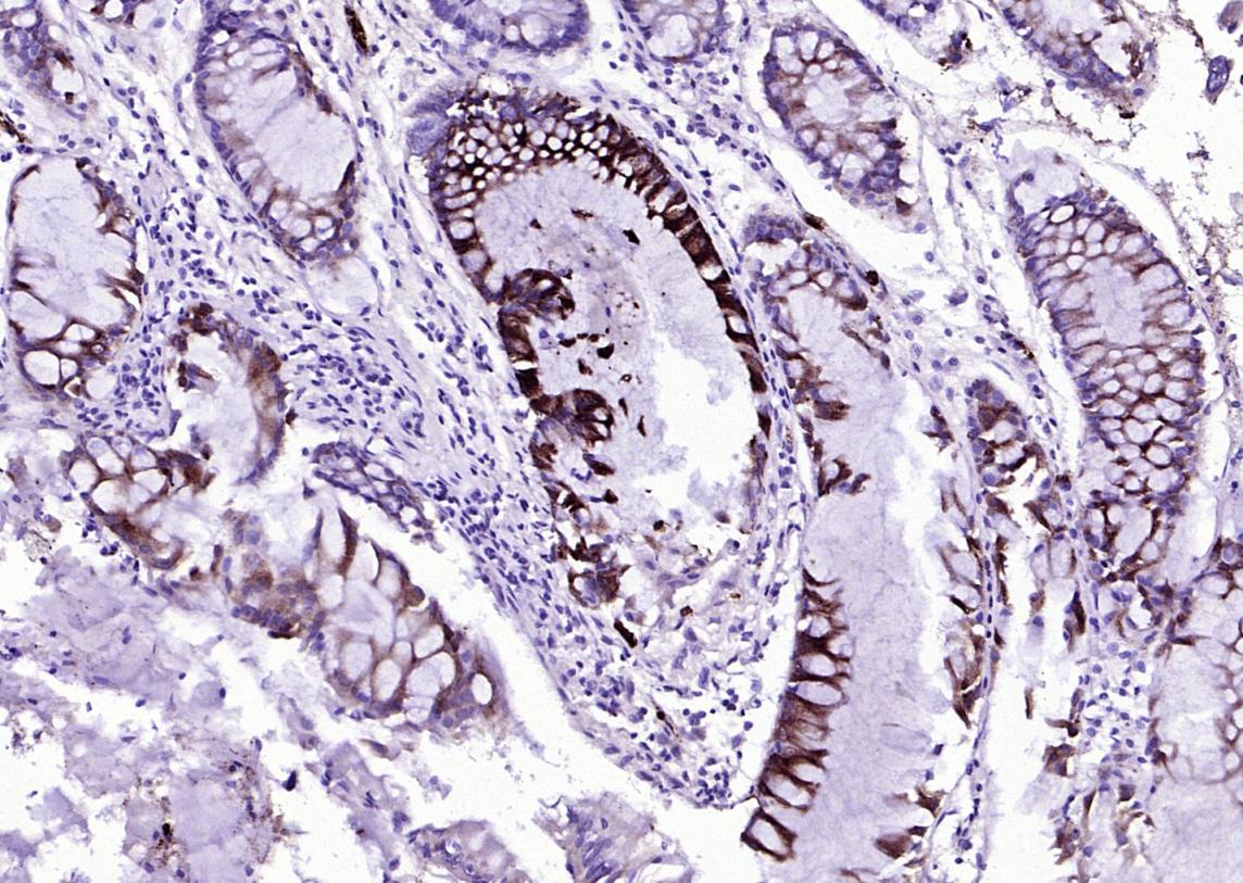
- 产品详情
- 实验流程
- 背景知识
Application 
| IHC-P, IHC-F, IF |
|---|---|
| Primary Accession | P80188 |
| Reactivity | Human |
| Host | Rabbit |
| Clonality | Polyclonal |
| Calculated MW | 22588 Da |
| Physical State | Liquid |
| Immunogen | KLH conjugated synthetic peptide derived from human Lipocalin 2 |
| Epitope Specificity | 101-198/198 |
| Isotype | IgG |
| Purity | affinity purified by Protein A |
| Buffer | 0.01M TBS (pH7.4) with 1% BSA, 0.02% Proclin300 and 50% Glycerol. |
| SUBCELLULAR LOCATION | Secreted. Note=Upon binding to the SLC22A17 (24p3R) receptor, it is internalized. |
| Important Note | This product as supplied is intended for research use only, not for use in human, therapeutic or diagnostic applications. |
| Background Descriptions | Lipocalin 2 is a member of the lipocalin family which encompass more than 25 members (including proteins like retinol binding protein, a1 microglobulin, b lactoglobulin, apolipoprotein D and odorant binding protein). The members of the lipocalin family are characterized by their ability to bind small lipophilic substances in their hydrophobic core, and thereby serve as transporters of substances. |
| Gene ID | 3934 |
|---|---|
| Other Names | Neutrophil gelatinase-associated lipocalin, NGAL, 25 kDa alpha-2-microglobulin-related subunit of MMP-9, Lipocalin-2, Oncogene 24p3, Siderocalin, p25, LCN2, HNL, NGAL {ECO:0000303|PubMed:8060329} |
| Dilution | IHC-P=1:100-500,IHC-F=1:100-500,IF=1:100-500,ELISA=1:5000-10000 |
| Storage | Store at -20 °C for one year. Avoid repeated freeze/thaw cycles. When reconstituted in sterile pH 7.4 0.01M PBS or diluent of antibody the antibody is stable for at least two weeks at 2-4 °C. |
For Research Use Only. Not For Use In Diagnostic Procedures.
| Name | LCN2 |
|---|---|
| Synonyms | HNL, NGAL {ECO:0000303|PubMed:8060329} |
| Function | Iron-trafficking protein involved in multiple processes such as apoptosis, innate immunity and renal development (PubMed:12453413, PubMed:20581821, PubMed:27780864). Binds iron through association with 2,3-dihydroxybenzoic acid (2,3-DHBA), a siderophore that shares structural similarities with bacterial enterobactin, and delivers or removes iron from the cell, depending on the context. Iron-bound form (holo-24p3) is internalized following binding to the SLC22A17 (24p3R) receptor, leading to release of iron and subsequent increase of intracellular iron concentration. In contrast, association of the iron- free form (apo-24p3) with the SLC22A17 (24p3R) receptor is followed by association with an intracellular siderophore, iron chelation and iron transfer to the extracellular medium, thereby reducing intracellular iron concentration. Involved in apoptosis due to interleukin-3 (IL3) deprivation: iron-loaded form increases intracellular iron concentration without promoting apoptosis, while iron-free form decreases intracellular iron levels, inducing expression of the proapoptotic protein BCL2L11/BIM, resulting in apoptosis (By similarity). Involved in innate immunity; limits bacterial proliferation by sequestering iron bound to microbial siderophores, such as enterobactin (PubMed:27780864). Can also bind siderophores from M.tuberculosis (PubMed:15642259, PubMed:21978368). |
| Cellular Location | Secreted. Cytoplasmic granule lumen. Cytoplasmic vesicle lumen. Note=Upon binding to the SLC22A17 (24p3R) receptor, it is internalized (By similarity). Releases the bound iron in the acidic lumen of cytoplasmic vesicles (PubMed:12453413, PubMed:20581821). {ECO:0000250|UniProtKB:P11672, ECO:0000269|PubMed:12453413, ECO:0000269|PubMed:20581821} |
| Tissue Location | Detected in neutrophils (at protein level) (PubMed:7683678, PubMed:8298140). Expressed in bone marrow and in tissues that are prone to exposure to microorganism (PubMed:9339356) High expression is found in bone marrow as well as in uterus, prostate, salivary gland, stomach, appendix, colon, trachea and lung (PubMed:9339356). Expressed in the medullary tubules of the kidney (PubMed:30418175). Not found in the small intestine or peripheral blood leukocytes (PubMed:9339356). |
Provided below are standard protocols that you may find useful for product applications.
BACKGROUND
This product as supplied is intended for research use only, not for use in human, therapeutic or diagnostic applications.
REFERENCES
Bundgaard J.R.,et al.Biochem. Biophys. Res. Commun. 202:1468-1475(1994).
Cowland J.B.,et al.Genomics 45:17-23(1997).
Ota T.,et al.Nat. Genet. 36:40-45(2004).
Ebert L.,et al.Submitted (JUN-2004) to the EMBL/GenBank/DDBJ databases.
Humphray S.J.,et al.Nature 429:369-374(2004).

终于等到您。ABCEPTA(百远生物)抗体产品。
点击下方“我要评价 ”按钮提交您的反馈信息,您的反馈和评价是我们最宝贵的财富之一,
我们将在1-3个工作日内处理您的反馈信息。
如有疑问,联系:0512-88856768 tech-china@abcepta.com.


