


 癌症的基本特征包括细胞增殖、血管生成、迁移、凋亡逃避机制和细胞永生等。找到癌症发生过程中这些通路的关键标记物和对应的抗体用于检测至关重要。
 为您推荐一个泛素化位点预测神器——泛素化分析工具,可以为您的蛋白的泛素化位点作出预测和评分。
 细胞自噬受体图形绘图工具为你的蛋白的细胞受体结合位点作出预测和评分,识别结合到自噬通路中的蛋白是非常重要的,便于让我们理解自噬在正常生理、病理过程中的作用,如发育、细胞分化、神经退化性疾病、压力条件下、感染和癌症。
Polycystin 1 Rabbit pAb
Polycystin 1 Rabbit pAb
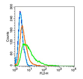
- 产品详情
- 实验流程
- 背景知识
| Primary Accession | P98161 |
|---|---|
| Reactivity | Human |
| Predicted | Mouse |
| Host | Rabbit |
| Clonality | Polyclonal |
| Calculated MW | 462529 Da |
| Physical State | Liquid |
| Immunogen | KLH conjugated synthetic peptide derived from human Polycystin 1 |
| Epitope Specificity | 131-230/4303 |
| Isotype | IgG |
| Purity | affinity purified by Protein A |
| Buffer | 0.01M TBS (pH7.4) with 1% BSA, 0.02% Proclin300 and 50% Glycerol. |
| SUBCELLULAR LOCATION | Membrane; Multi-pass membrane protein. Cell projection, cilium. Note=PKD1 localization to the plasma and ciliary membranes requires PKD2, is independent of PKD2 channel activity, and involves stimulation of PKD1 autoproteolytic cleavage at the GPS domain. |
| DISEASE | Defects in PKD1 are the cause of polycystic kidney disease autosomal dominant type 1 (ADPKD1) [MIM:173900]. ADPKD is characterized by progressive formation and enlargement of cysts in both kidneys, typically leading to end-stage renal disease in adult life. Cysts also occurs in the liver and other organs. Its prevalence is estimated at about 1/1000. |
| Important Note | This product as supplied is intended for research use only, not for use in human, therapeutic or diagnostic applications. |
| Background Descriptions | This gene encodes a member of the polycystin protein family. The encoded glycoprotein contains a large N-terminal extracellular region, multiple transmembrane domains and a cytoplasmic C-tail. It is an integral membrane protein that functions as a regulator of calcium permeable cation channels and intracellular calcium homoeostasis. It is also involved in cell-cell/matrix interactions and may modulate G-protein-coupled signal-transduction pathways. It plays a role in renal tubular development, and mutations in this gene cause autosomal dominant polycystic kidney disease type 1 (ADPKD1). ADPKD1 is characterized by the growth of fluid-filled cysts that replace normal renal tissue and result in end-stage renal failure. Splice variants encoding different isoforms have been noted for this gene. Also, six pseudogenes, closely linked in a known duplicated region on chromosome 16p, have been described. [provided by RefSeq]. |
| Gene ID | 5310 |
|---|---|
| Other Names | Polycystin-1, PC1, Autosomal dominant polycystic kidney disease 1 protein, PKD1 (HGNC:9008) |
| Dilution | Flow-Cyt=1 µg/Test |
| Storage | Store at -20 °C for one year. Avoid repeated freeze/thaw cycles. When reconstituted in sterile pH 7.4 0.01M PBS or diluent of antibody the antibody is stable for at least two weeks at 2-4 °C. |
For Research Use Only. Not For Use In Diagnostic Procedures.
| Name | PKD1 (HGNC:9008) |
|---|---|
| Function | Component of a heteromeric calcium-permeable ion channel formed by PKD1 and PKD2 that is activated by interaction between PKD1 and a Wnt family member, such as WNT3A and WNT9B (PubMed:27214281). Both PKD1 and PKD2 are required for channel activity (PubMed:27214281). Involved in renal tubulogenesis (PubMed:12482949). Involved in fluid- flow mechanosensation by the primary cilium in renal epithelium (By similarity). Acts as a regulator of cilium length, together with PKD2 (By similarity). The dynamic control of cilium length is essential in the regulation of mechanotransductive signaling (By similarity). The cilium length response creates a negative feedback loop whereby fluid shear-mediated deflection of the primary cilium, which decreases intracellular cAMP, leads to cilium shortening and thus decreases flow- induced signaling (By similarity). May be an ion-channel regulator. Involved in adhesive protein-protein and protein-carbohydrate interactions. Likely to be involved with polycystin-1-interacting protein 1 in the detection, sequestration and exocytosis of senescent mitochondria (PubMed:37681898). |
| Cellular Location | Cell membrane; Multi-pass membrane protein. Cell projection, cilium {ECO:0000250|UniProtKB:O08852}. Endoplasmic reticulum {ECO:0000250|UniProtKB:O08852}. Golgi apparatus {ECO:0000250|UniProtKB:O08852}. Vesicle Secreted, extracellular exosome Note=PKD1 localization to the plasma and ciliary membranes requires PKD2, is independent of PKD2 channel activity, and involves stimulation of PKD1 autoproteolytic cleavage at the GPS region of the GAIN-B domain. PKD1:PKD2 interaction is required to reach the Golgi apparatus from endoplasmic reticulum and then traffic to the cilia (By similarity). Ciliary localization of PKD1 requires BBS1 and ARL6/BBS3 (By similarity). Cell surface localization requires GANAB (PubMed:27259053). Detected on migrasomes and on extracellular exosomes in urine (PubMed:37681898). {ECO:0000250|UniProtKB:O08852, ECO:0000269|PubMed:27259053, ECO:0000269|PubMed:37681898} |
Research Areas
Application Protocols
Provided below are standard protocols that you may find useful for product applications.
BACKGROUND
This product as supplied is intended for research use only, not for use in human, therapeutic or diagnostic applications.
REFERENCES
Gluecksmann-Kuis M.A.,et al.Cell 81:289-298(1995).
Hughes J.,et al.Nat. Genet. 10:151-160(1995).
Martin J.,et al.Nature 432:988-994(2004).
Ward C.J.,et al.Cell 77:881-894(1994).
Ward C.J.,et al.Cell 78:725-725(1994).

终于等到您。ABCEPTA(百远生物)抗体产品。
点击下方“我要评价 ”按钮提交您的反馈信息,您的反馈和评价是我们最宝贵的财富之一,
我们将在1-3个工作日内处理您的反馈信息。
如有疑问,联系:0512-88856768 tech-china@abcepta.com.


