


 癌症的基本特征包括细胞增殖、血管生成、迁移、凋亡逃避机制和细胞永生等。找到癌症发生过程中这些通路的关键标记物和对应的抗体用于检测至关重要。
 为您推荐一个泛素化位点预测神器——泛素化分析工具,可以为您的蛋白的泛素化位点作出预测和评分。
 细胞自噬受体图形绘图工具为你的蛋白的细胞受体结合位点作出预测和评分,识别结合到自噬通路中的蛋白是非常重要的,便于让我们理解自噬在正常生理、病理过程中的作用,如发育、细胞分化、神经退化性疾病、压力条件下、感染和癌症。
ROCK1 Rabbit pAb
ROCK1 Rabbit pAb
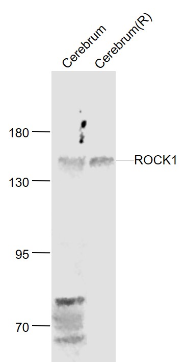
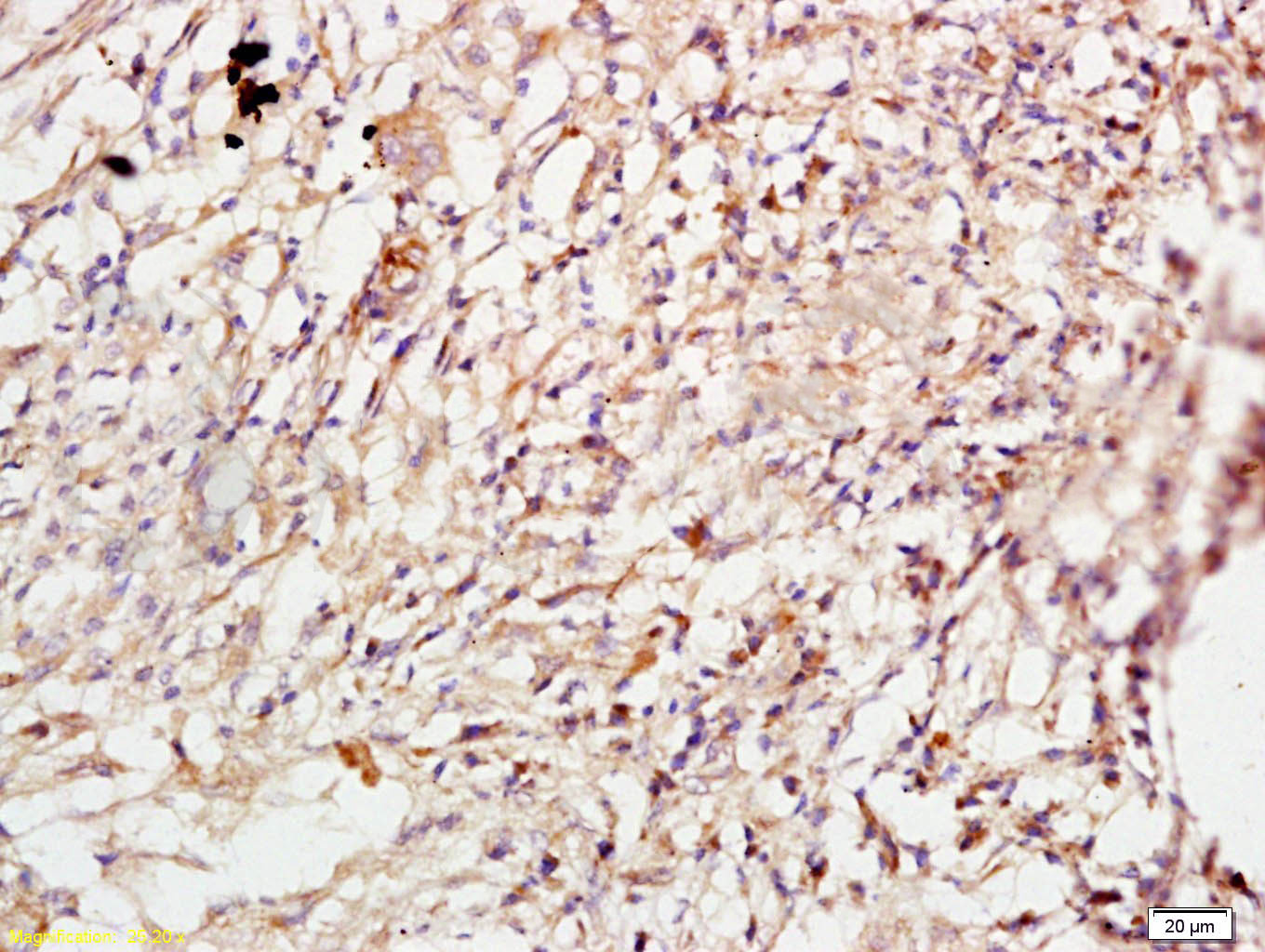
- 产品详情
- 实验流程
- 背景知识
Application 
| WB, IHC-P, IHC-F, IF |
|---|---|
| Primary Accession | Q13464 |
| Reactivity | Rat, Human, Mouse |
| Predicted | Rabbit |
| Host | Rabbit |
| Clonality | Polyclonal |
| Calculated MW | 158175 Da |
| Physical State | Liquid |
| Immunogen | KLH conjugated synthetic peptide derived from human ROCK1 |
| Epitope Specificity | 1001-1200/1354 |
| Isotype | IgG |
| Purity | affinity purified by Protein A |
| Buffer | 0.01M TBS (pH7.4) with 1% BSA, 0.02% Proclin300 and 50% Glycerol. |
| SUBCELLULAR LOCATION | Cytoplasm. Cytoplasm, cytoskeleton, centrosome, centriole (By similarity). Golgi apparatus membrane; Peripheral membrane protein. Cell projection, bleb. Note=Associated with the mother centriole and an intercentriolar linker (By similarity). A small proportion is associated with Golgi membranes. |
| DISEASE | Defects in SOD1 are the cause of amyotrophic lateral sclerosis type 1 (ALS1) [MIM:105400]. ALS1 is a familial form of amyotrophic lateral sclerosis, a neurodegenerative disorder affecting upper and lower motor neurons and resulting in fatal paralysis. Sensory abnormalities are absent. Death usually occurs within 2 to 5 years. The etiology of amyotrophic lateral sclerosis is likely to be multifactorial, involving both genetic and environmental factors. The disease is inherited in 5-10% of cases leading to familial forms. |
| Important Note | This product as supplied is intended for research use only, not for use in human, therapeutic or diagnostic applications. |
| Background Descriptions | This gene encodes a protein serine/threonine kinase that is activated when bound to the GTP-bound form of Rho. The small GTPase Rho regulates formation of focal adhesions and stress fibers of fibroblasts, as well as adhesion and aggregation of platelets and lymphocytes by shuttling between the inactive GDP-bound form and the active GTP-bound form. Rho is also essential in cytokinesis and plays a role in transcriptional activation by serum response factor. This protein, a downstream effector of Rho, phosphorylates and activates LIM kinase, which in turn, phosphorylates cofilin, inhibiting its actin-depolymerizing activity. [provided by RefSeq]. |
| Gene ID | 6093 |
|---|---|
| Other Names | Rho-associated protein kinase 1, 2.7.11.39, Renal carcinoma antigen NY-REN-35, Rho-associated, coiled-coil-containing protein kinase 1, Rho-associated, coiled-coil-containing protein kinase I, ROCK-I, p160 ROCK-1, p160ROCK, ROCK1 |
| Dilution | WB=1:500-2000,IHC-P=1:100-500,IHC-F=1:100-500,IF=1:100-500 |
| Storage | Store at -20 °C for one year. Avoid repeated freeze/thaw cycles. When reconstituted in sterile pH 7.4 0.01M PBS or diluent of antibody the antibody is stable for at least two weeks at 2-4 °C. |
For Research Use Only. Not For Use In Diagnostic Procedures.
| Name | ROCK1 |
|---|---|
| Function | Protein kinase which is a key regulator of the actin cytoskeleton and cell polarity (PubMed:10436159, PubMed:10652353, PubMed:11018042, PubMed:11283607, PubMed:17158456, PubMed:18573880, PubMed:19131646, PubMed:8617235, PubMed:9722579). Involved in regulation of smooth muscle contraction, actin cytoskeleton organization, stress fiber and focal adhesion formation, neurite retraction, cell adhesion and motility via phosphorylation of DAPK3, GFAP, LIMK1, LIMK2, MYL9/MLC2, TPPP, PFN1 and PPP1R12A (PubMed:10436159, PubMed:10652353, PubMed:11018042, PubMed:11283607, PubMed:17158456, PubMed:18573880, PubMed:19131646, PubMed:23093407, PubMed:23355470, PubMed:8617235, PubMed:9722579). Phosphorylates FHOD1 and acts synergistically with it to promote SRC-dependent non-apoptotic plasma membrane blebbing (PubMed:18694941). Phosphorylates JIP3 and regulates the recruitment of JNK to JIP3 upon UVB-induced stress (PubMed:19036714). Acts as a suppressor of inflammatory cell migration by regulating PTEN phosphorylation and stability (By similarity). Acts as a negative regulator of VEGF-induced angiogenic endothelial cell activation (PubMed:19181962). Required for centrosome positioning and centrosome-dependent exit from mitosis (By similarity). Plays a role in terminal erythroid differentiation (PubMed:21072057). Inhibits podocyte motility via regulation of actin cytoskeletal dynamics and phosphorylation of CFL1 (By similarity). Promotes keratinocyte terminal differentiation (PubMed:19997641). Involved in osteoblast compaction through the fibronectin fibrillogenesis cell-mediated matrix assembly process, essential for osteoblast mineralization (By similarity). May regulate closure of the eyelids and ventral body wall by inducing the assembly of actomyosin bundles (By similarity). |
| Cellular Location | Cytoplasm. Cytoplasm, cytoskeleton, microtubule organizing center, centrosome, centriole {ECO:0000250|UniProtKB:P70335}. Golgi apparatus membrane; Peripheral membrane protein. Cell projection, bleb. Cytoplasm, cytoskeleton {ECO:0000250|UniProtKB:P70335}. Cell membrane {ECO:0000250|UniProtKB:P70335}. Cell projection, lamellipodium {ECO:0000250|UniProtKB:P70335}. Cell projection, ruffle {ECO:0000250|UniProtKB:P70335}. Note=A small proportion is associated with Golgi membranes (PubMed:12773565). Associated with the mother centriole and an intercentriolar linker (By similarity). Colocalizes with ITGB1BP1 and ITGB1 at the cell membrane predominantly in lamellipodia and membrane ruffles, but also in retraction fibers (By similarity). Localizes at the cell membrane in an ITGB1BP1-dependent manner (By similarity). {ECO:0000250|UniProtKB:P70335, ECO:0000269|PubMed:12773565} |
| Tissue Location | Detected in blood platelets. |
Provided below are standard protocols that you may find useful for product applications.
BACKGROUND
This product as supplied is intended for research use only, not for use in human, therapeutic or diagnostic applications.
REFERENCES
Ishizaki T.,et al.EMBO J. 15:1885-1893(1996).
Bienvenut W.V.,et al.Submitted (MAR-2009) to UniProtKB.
Totoki Y.,et al.Submitted (MAR-2005) to the EMBL/GenBank/DDBJ databases.
Van Eyk J.E.,et al.J. Biol. Chem. 273:23433-23439(1998).
Scanlan M.J.,et al.Int. J. Cancer 83:456-464(1999).

终于等到您。ABCEPTA(百远生物)抗体产品。
点击下方“我要评价 ”按钮提交您的反馈信息,您的反馈和评价是我们最宝贵的财富之一,
我们将在1-3个工作日内处理您的反馈信息。
如有疑问,联系:0512-88856768 tech-china@abcepta.com.


