


 癌症的基本特征包括细胞增殖、血管生成、迁移、凋亡逃避机制和细胞永生等。找到癌症发生过程中这些通路的关键标记物和对应的抗体用于检测至关重要。
 为您推荐一个泛素化位点预测神器——泛素化分析工具,可以为您的蛋白的泛素化位点作出预测和评分。
 细胞自噬受体图形绘图工具为你的蛋白的细胞受体结合位点作出预测和评分,识别结合到自噬通路中的蛋白是非常重要的,便于让我们理解自噬在正常生理、病理过程中的作用,如发育、细胞分化、神经退化性疾病、压力条件下、感染和癌症。
XIAP/BIRC4 Rabbit pAb
XIAP/BIRC4 Rabbit pAb
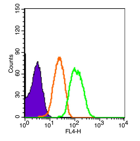
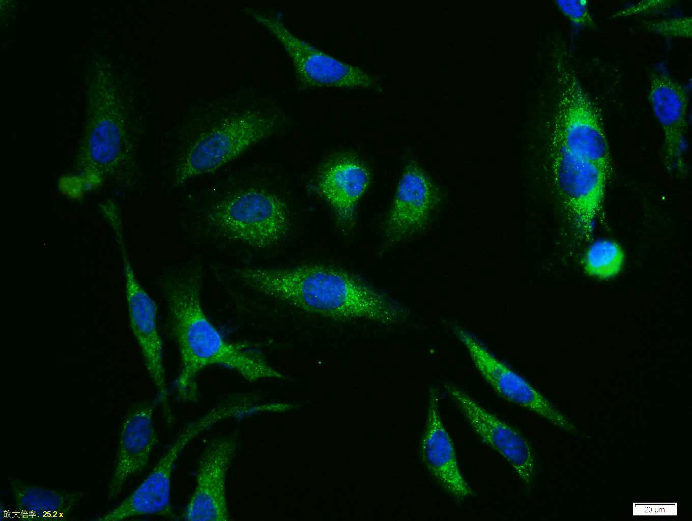
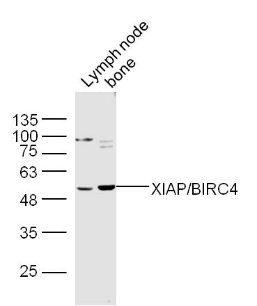
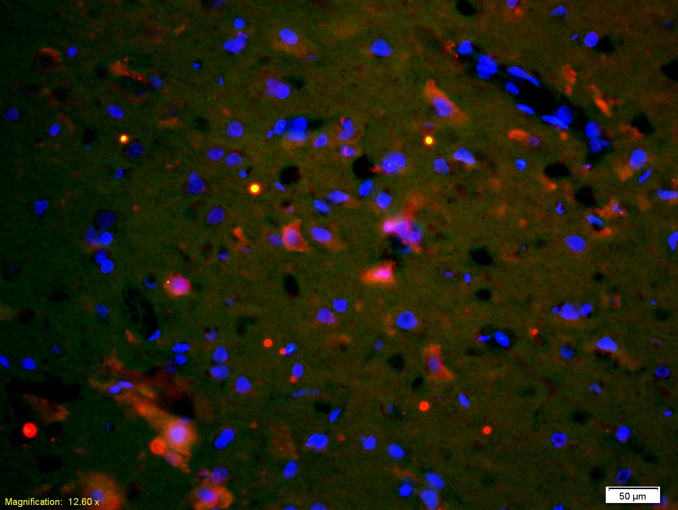
- 产品详情
- 实验流程
- 背景知识
Application 
| WB, IHC-P, IHC-F, IF |
|---|---|
| Primary Accession | P98170 |
| Reactivity | Rat, Human, Mouse |
| Host | Rabbit |
| Clonality | Polyclonal |
| Calculated MW | 56685 Da |
| Physical State | Liquid |
| Immunogen | KLH conjugated synthetic peptide derived from human XIAP |
| Epitope Specificity | 201-330/496 |
| Isotype | IgG |
| Purity | affinity purified by Protein A |
| Buffer | 0.01M TBS (pH7.4) with 1% BSA, 0.02% Proclin300 and 50% Glycerol. |
| SUBCELLULAR LOCATION | Cytoplasm. |
| DISEASE | Defects in XIAP are the cause of lymphoproliferative syndrome X-linked type 2 (XLP2) [MIM:300635]. XLP is a rare immunodeficiency characterized by extreme susceptibility to infection with Epstein-Barr virus (EBV). Symptoms include severe or fatal mononucleosis, acquired hypogammaglobulinemia, pancytopenia and malignant lymphoma. |
| Important Note | This product as supplied is intended for research use only, not for use in human, therapeutic or diagnostic applications. |
| Background Descriptions | This gene encodes a protein that belongs to a family of apoptotic suppressor proteins. Members of this family share a conserved motif termed, baculovirus IAP repeat, which is necessary for their anti-apoptotic function. This protein functions through binding to tumor necrosis factor receptor-associated factors TRAF1 and TRAF2 and inhibits apoptosis induced by menadione, a potent inducer of free radicals, and interleukin 1-beta converting enzyme. This protein also inhibits at least two members of the caspase family of cell-death proteases, caspase-3 and caspase-7. Mutations in this gene are the cause of X-linked lymphoproliferative syndrome. Alternate splicing results in multiple transcript variants. Pseudogenes of this gene are found on chromosomes 2 and 11.[provided by RefSeq, Feb 2011] |
| Gene ID | 331 |
|---|---|
| Other Names | E3 ubiquitin-protein ligase XIAP, 2.3.2.27, Baculoviral IAP repeat-containing protein 4, IAP-like protein, ILP, hILP, Inhibitor of apoptosis protein 3, IAP-3, hIAP-3, hIAP3, RING-type E3 ubiquitin transferase XIAP, X-linked inhibitor of apoptosis protein, X-linked IAP, XIAP {ECO:0000303|PubMed:12121969, ECO:0000312|HGNC:HGNC:592} |
| Dilution | WB=1:500-2000,IHC-P=1:100-500,IHC-F=1:100-500,ICC/IF=1:100-500,IF=1:100-500,Flow-Cyt=3ug/Test |
| Storage | Store at -20 °C for one year. Avoid repeated freeze/thaw cycles. When reconstituted in sterile pH 7.4 0.01M PBS or diluent of antibody the antibody is stable for at least two weeks at 2-4 °C. |
For Research Use Only. Not For Use In Diagnostic Procedures.
| Name | XIAP {ECO:0000303|PubMed:12121969, ECO:0000312|HGNC:HGNC:592} |
|---|---|
| Function | Multi-functional protein which regulates not only caspases and apoptosis, but also modulates inflammatory signaling and immunity, copper homeostasis, mitogenic kinase signaling, cell proliferation, as well as cell invasion and metastasis (PubMed:11257230, PubMed:11257231, PubMed:11447297, PubMed:12121969, PubMed:12620238, PubMed:17560374, PubMed:17967870, PubMed:19473982, PubMed:20154138, PubMed:22103349, PubMed:9230442). Acts as a direct caspase inhibitor (PubMed:11257230, PubMed:11257231, PubMed:12620238). Directly bind to the active site pocket of CASP3 and CASP7 and obstructs substrate entry (PubMed:11257230, PubMed:11257231, PubMed:16352606, PubMed:16916640). Inactivates CASP9 by keeping it in a monomeric, inactive state (PubMed:12620238). Acts as an E3 ubiquitin-protein ligase regulating NF-kappa-B signaling and the target proteins for its E3 ubiquitin- protein ligase activity include: RIPK1, RIPK2, MAP3K2/MEKK2, DIABLO/SMAC, AIFM1, CCS, PTEN and BIRC5/survivin (PubMed:17560374, PubMed:17967870, PubMed:19473982, PubMed:20154138, PubMed:22103349, PubMed:22607974, PubMed:29452636, PubMed:30026309). Acts as an important regulator of innate immunity by mediating 'Lys-63'-linked polyubiquitination of RIPK2 downstream of NOD1 and NOD2, thereby transforming RIPK2 into a scaffolding protein for downstream effectors, ultimately leading to activation of the NF-kappa-B and MAP kinases signaling (PubMed:19667203, PubMed:22607974, PubMed:29452636, PubMed:30026309). 'Lys-63'-linked polyubiquitination of RIPK2 also promotes recruitment of the LUBAC complex to RIPK2 (PubMed:22607974, PubMed:29452636). Regulates the BMP signaling pathway and the SMAD and MAP3K7/TAK1 dependent pathways leading to NF-kappa-B and JNK activation (PubMed:17560374). Ubiquitination of CCS leads to enhancement of its chaperone activity toward its physiologic target, SOD1, rather than proteasomal degradation (PubMed:20154138). Ubiquitination of MAP3K2/MEKK2 and AIFM1 does not lead to proteasomal degradation (PubMed:17967870, PubMed:22103349). Plays a role in copper homeostasis by ubiquitinating COMMD1 and promoting its proteasomal degradation (PubMed:14685266). Can also function as E3 ubiquitin-protein ligase of the NEDD8 conjugation pathway, targeting effector caspases for neddylation and inactivation (PubMed:21145488). Ubiquitinates and therefore mediates the proteasomal degradation of BCL2 in response to apoptosis (PubMed:29020630). Protects cells from spontaneous formation of the ripoptosome, a large multi-protein complex that has the capability to kill cancer cells in a caspase-dependent and caspase- independent manner (PubMed:22095281). Suppresses ripoptosome formation by ubiquitinating RIPK1 and CASP8 (PubMed:22095281). Acts as a positive regulator of Wnt signaling and ubiquitinates TLE1, TLE2, TLE3, TLE4 and AES (PubMed:22304967). Ubiquitination of TLE3 results in inhibition of its interaction with TCF7L2/TCF4 thereby allowing efficient recruitment and binding of the transcriptional coactivator beta-catenin to TCF7L2/TCF4 that is required to initiate a Wnt-specific transcriptional program (PubMed:22304967). |
| Cellular Location | Cytoplasm. Nucleus. Note=TLE3 promotes its nuclear localization. |
| Tissue Location | Expressed in colonic crypts (at protein level) (PubMed:30389919). Ubiquitous, except peripheral blood leukocytes (PubMed:8654366). |
Provided below are standard protocols that you may find useful for product applications.
BACKGROUND
This product as supplied is intended for research use only, not for use in human, therapeutic or diagnostic applications.
REFERENCES
Uren A.G.,et al.Proc. Natl. Acad. Sci. U.S.A. 93:4974-4978(1996).
Farahani R.,et al.Submitted (JUN-1997) to the EMBL/GenBank/DDBJ databases.
Church D.M.,et al.PLoS Biol. 7:E1000112-E1000112(2009).
Mural R.J.,et al.Submitted (JUL-2005) to the EMBL/GenBank/DDBJ databases.
Winsauer G.,et al.Cell. Signal. 20:2107-2112(2008).

终于等到您。ABCEPTA(百远生物)抗体产品。
点击下方“我要评价 ”按钮提交您的反馈信息,您的反馈和评价是我们最宝贵的财富之一,
我们将在1-3个工作日内处理您的反馈信息。
如有疑问,联系:0512-88856768 tech-china@abcepta.com.


