


 癌症的基本特征包括细胞增殖、血管生成、迁移、凋亡逃避机制和细胞永生等。找到癌症发生过程中这些通路的关键标记物和对应的抗体用于检测至关重要。
 为您推荐一个泛素化位点预测神器——泛素化分析工具,可以为您的蛋白的泛素化位点作出预测和评分。
 细胞自噬受体图形绘图工具为你的蛋白的细胞受体结合位点作出预测和评分,识别结合到自噬通路中的蛋白是非常重要的,便于让我们理解自噬在正常生理、病理过程中的作用,如发育、细胞分化、神经退化性疾病、压力条件下、感染和癌症。
RAGE Rabbit pAb
RAGE Rabbit pAb
- 产品详情
- 实验流程
- 背景知识
Application 
| WB, IHC-P, IHC-F, IF |
|---|---|
| Primary Accession | Q63495 |
| Reactivity | Rat, Human, Mouse |
| Host | Rabbit |
| Clonality | Polyclonal |
| Calculated MW | 42664 Da |
| Physical State | Liquid |
| Immunogen | KLH conjugated synthetic peptide derived from rat AGER |
| Epitope Specificity | 151-250/403 |
| Isotype | IgG |
| Purity | affinity purified by Protein A |
| Buffer | 0.01M TBS (pH7.4) with 1% BSA, 0.02% Proclin300 and 50% Glycerol. |
| SUBCELLULAR LOCATION | Isoform 1: Cell membrane; Single-pass type I membrane protein. Isoform 2: Secreted. |
| Important Note | This product as supplied is intended for research use only, not for use in human, therapeutic or diagnostic applications. |
| Background Descriptions | Advanced glycosylation end product-specific receptor (AGER; RAGE) is a member of the immunoglobulin superfamily of cell surface molecules that binds molecules that have been irreversibly modified by non-enzymatic glycation and oxidation, and are know as advanced glycation end products (AGEs). It is expressed by endothelium, mononuclear phagocytes, neurons and smooth muscle cells. Whereas RAGE is present at high levels during development, especially in the central nervous system, its levels decline during maturity.The increased expression of RAGE is associated with several pathological states, such as diabetic vasculopathy, neuropathy, retinopathy and other disorders, including Alzheimer's disease and immune/inflammatory reactions of the vessel walls. In diabetic tissues, the production of RAGE is due to the overproduction of AGEs that eventually overwhelm the protective properties of RAGE. This results in oxidative stress and endothelial cell dysfunction that leads to vascular disease in diabetics. In the brain, RAGE also binds amyloid beta (Ab). Because Ab is overproduced in neurons and vessels in the brains of Alzheimer disease, this leads to the hyperstimulation of RAGE. The RAGE-Ab interaction is thought to result in oxidative stress leading to neuronal degeneration. |
| Other Names | Advanced glycosylation end product-specific receptor, Receptor for advanced glycosylation end products, Ager, Rage |
|---|---|
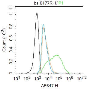
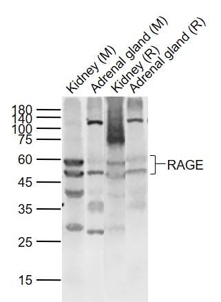
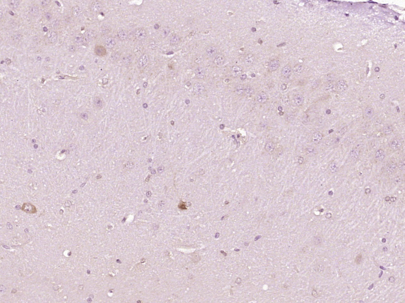
| Dilution | WB=1:500-2000,IHC-P=1:100-500,IHC-F=1:100-500,IF=1:100-500,Flow-Cyt=1 µg /test |
| Storage | Store at -20 °C for one year. Avoid repeated freeze/thaw cycles. When reconstituted in sterile pH 7.4 0.01M PBS or diluent of antibody the antibody is stable for at least two weeks at 2-4 °C. |
For Research Use Only. Not For Use In Diagnostic Procedures.
| Name | Ager |
|---|---|
| Synonyms | Rage |
| Function | Cell surface pattern recognition receptor that senses endogenous stress signals with a broad ligand repertoire including advanced glycation end products, S100 proteins, high-mobility group box 1 protein/HMGB1, amyloid beta/APP oligomers, nucleic acids, histones, phospholipids and glycosaminoglycans (PubMed:19910580, PubMed:28627626). Advanced glycation end products (AGEs) are nonenzymatically glycosylated proteins which accumulate in vascular tissue in aging and at an accelerated rate in diabetes. These ligands accumulate at inflammatory sites during the pathogenesis of various diseases including diabetes, vascular complications, neurodegenerative disorders and cancers, and RAGE transduces their binding into pro- inflammatory responses. Upon ligand binding, uses TIRAP and MYD88 as adapters to transduce the signal ultimately leading to the induction of inflammatory cytokines IL6, IL8 and TNFalpha through activation of NF- kappa-B. Interaction with S100A12 on endothelium, mononuclear phagocytes, and lymphocytes triggers cellular activation, with generation of key pro-inflammatory mediators (By similarity). Interaction with S100B after myocardial infarction may play a role in myocyte apoptosis by activating ERK1/2 and p53/TP53 signaling (By similarity). Contributes to the translocation of amyloid-beta peptide (ABPP) across the cell membrane from the extracellular to the intracellular space in cortical neurons. ABPP-initiated RAGE signaling, especially stimulation of p38 mitogen-activated protein kinase (MAPK), has the capacity to drive a transport system delivering ABPP as a complex with RAGE to the intraneuronal space. Participates in endothelial albumin transcytosis together with HMGB1 through the RAGE/SRC/Caveolin-1 pathway, leading to endothelial hyperpermeability. Mediates the loading of HMGB1 in extracellular vesicles (EVs) that shuttle HMGB1 to hepatocytes by transferrin-mediated endocytosis and subsequently promote hepatocyte pyroptosis by activating the NLRP3 inflammasome. Binds to DNA and promotes extracellular hypomethylated DNA (CpG DNA) uptake by cells via the endosomal route to activate inflammatory responses (By similarity). Mediates phagocytosis by non- professional phagocytes (NPP) and this is enhanced by binding to ligands including RNA, DNA, HMGB1 and histones (By similarity). Promotes NPP-mediated phagocytosis of Saccharomyces cerevisiae spores by binding to RNA attached to the spore wall (By similarity). Also promotes NPP-mediated phagocytosis of apoptotic cells (By similarity). Following DNA damage, recruited to DNA double-strand break sites where it colocalizes with the MRN repair complex via interaction with double- strand break repair protein MRE11 (By similarity). Enhances the endonuclease activity of MRE11, promoting the end resection of damaged DNA (By similarity). Promotes DNA damage repair in trophoblasts which enhances trophoblast invasion and contributes to placental development and maintenance (By similarity). Protects cells from DNA replication stress by localizing to damaged replication forks where it stabilizes the MCM2-7 complex and promotes faithful progression of the replication fork (By similarity). |
| Cellular Location | Cell membrane; Single-pass type I membrane protein. Cell projection, phagocytic cup {ECO:0000250|UniProtKB:Q15109}. Early endosome {ECO:0000250|UniProtKB:Q15109}. Nucleus {ECO:0000250|UniProtKB:Q62151} Note=Nuclear translocation is enhanced by irradiation, hypoxia and reperfusion injury to brain or kidney. {ECO:0000250|UniProtKB:Q62151} |
| Tissue Location | Endothelial cells and cardiomyocytes (PubMed:19910580). Expressed in brain (PubMed:28627626) |
Research Areas
Application Protocols
Provided below are standard protocols that you may find useful for product applications.
BACKGROUND
This product as supplied is intended for research use only, not for use in human, therapeutic or diagnostic applications.
REFERENCES
Renard C.,et al.Mol. Pharmacol. 52:54-62(1997).
Tsoporis J.N.,et al.Circ. Res. 106:93-101(2010).

终于等到您。ABCEPTA(百远生物)抗体产品。
点击下方“我要评价 ”按钮提交您的反馈信息,您的反馈和评价是我们最宝贵的财富之一,
我们将在1-3个工作日内处理您的反馈信息。
如有疑问,联系:0512-88856768 tech-china@abcepta.com.


