


 癌症的基本特征包括细胞增殖、血管生成、迁移、凋亡逃避机制和细胞永生等。找到癌症发生过程中这些通路的关键标记物和对应的抗体用于检测至关重要。
 为您推荐一个泛素化位点预测神器——泛素化分析工具,可以为您的蛋白的泛素化位点作出预测和评分。
 细胞自噬受体图形绘图工具为你的蛋白的细胞受体结合位点作出预测和评分,识别结合到自噬通路中的蛋白是非常重要的,便于让我们理解自噬在正常生理、病理过程中的作用,如发育、细胞分化、神经退化性疾病、压力条件下、感染和癌症。
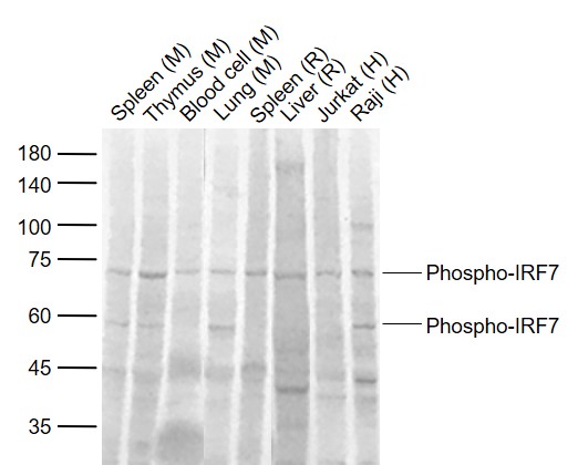
phospho-IRF7 (Ser471 + Ser472) Rabbit pAb
phospho-IRF7 (Ser471 + Ser472) Rabbit pAb
- 产品详情
- 实验流程
- 背景知识
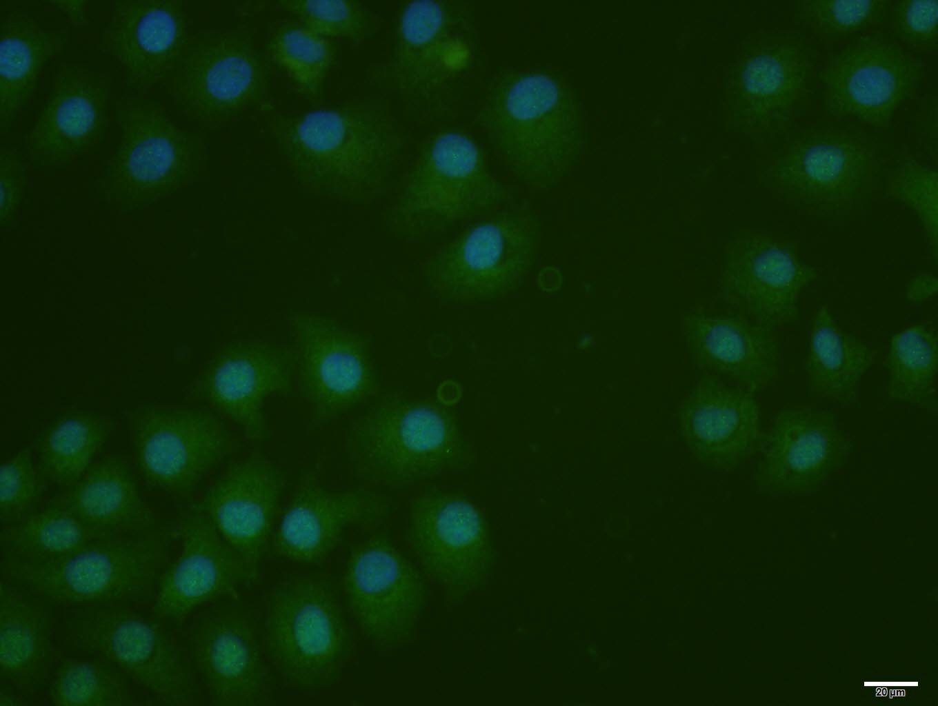
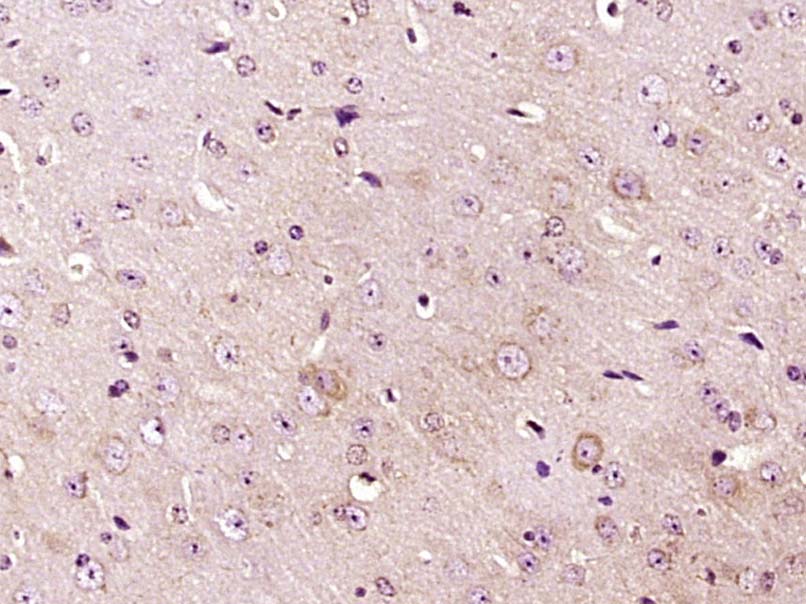
Application
| WB, IHC-P, IHC-F, IF |
|---|---|
| Primary Accession | Q92985 |
| Reactivity | Human, Mouse, Rat |
| Predicted | Pig, Horse |
| Host | Rabbit |
| Clonality | Polyclonal |
| Calculated MW | 54278 Da |
| Physical State | Liquid |
| Immunogen | KLH conjugated synthesised phosphopeptide derived from human IRF7 around the phosphorylation site of Ser471/472 |
| Epitope Specificity | GV(p-S)(p-S)LD |
| Isotype | IgG |
| Purity | affinity purified by Protein A |
| Buffer | 0.01M TBS (pH7.4) with 1% BSA, 0.02% Proclin300 and 50% Glycerol. |
| SUBCELLULAR LOCATION | Nucleus. Cytoplasm. Note=The phosphorylated and active form accumulates selectively in the nucleus. |
| SIMILARITY | Belongs to the IRF family.Contains 1 IRF tryptophan pentad repeat DNA-binding domain. |
| SUBUNIT | Monomer. Homodimer; phosphorylation-induced. Heterodimer with IRF3. Interacts with TICAM1 and TICAM2. Interacts with rotavirus A NSP1; this interaction leads to the proteasome-dependent degradation of IRF7. Interacts with Epstein-Barr virus LF2. Interacts with MYD88 AND TRAF6. |
| Post-translational modifications | Acetylation inhibits its DNA-binding ability and activity. In response to a viral infection, phosphorylated on Ser-477 and Ser-479 by TBK1 and IKBKE1. Phosphorylation, and subsequent activation is inhibited by vaccinia virus protein E3. In TLR7- and TLR9-mediated signaling pathway, phosphorylated by IRAK1.TRAF6-mediated ubiquitination is required for IRF7 activation (By similarity). Sumoylated by TRIM28, which inhibits its transactivation activity. |
| Important Note | This product as supplied is intended for research use only, not for use in human, therapeutic or diagnostic applications. |
| Background Descriptions | IRF7 encodes interferon regulatory factor 7, a member of the interferon regulatory transcription factor (IRF) family. IRF7 has been shown to play a role in the transcriptional activation of virus-inducible cellular genes, including interferon beta chain genes. Inducible expression of IRF7 is largely restricted to lymphoid tissue. Multiple IRF7 transcript variants have been identified, although the functional consequences of these have not yet been established. [provided by RefSeq, Jul 2008] |
| Gene ID | 3665 |
|---|---|
| Other Names | Interferon regulatory factor 7, IRF-7, IRF7 |
| Target/Specificity | Expressed predominantly in spleen, thymus and peripheral blood leukocytes. |
| Dilution | WB=1:500-2000,IHC-P=1:100-500,IHC-F=1:100-500,ICC/IF=1:100-500,IF=1:100-500 |
| Storage | Store at -20 °C for one year. Avoid repeated freeze/thaw cycles. When reconstituted in sterile pH 7.4 0.01M PBS or diluent of antibody the antibody is stable for at least two weeks at 2-4 °C. |
| Name | IRF7 |
|---|---|
| Function | Key transcriptional regulator of type I interferon (IFN)- dependent immune responses and plays a critical role in the innate immune response against DNA and RNA viruses (PubMed:28342865, PubMed:28768858). Regulates the transcription of type I IFN genes (IFN- alpha and IFN-beta) and IFN-stimulated genes (ISG) by binding to an interferon-stimulated response element (ISRE) in their promoters (PubMed:17574024, PubMed:32972995). Can efficiently activate both the IFN-beta (IFNB) and the IFN-alpha (IFNA) genes and mediate their induction via both the virus-activated, MyD88-independent pathway and the TLR-activated, MyD88-dependent pathway. Induces transcription of ubiquitin hydrolase USP25 mRNA in response to lipopolysaccharide (LPS) or viral infection in a type I IFN-dependent manner (By similarity). Required during both the early and late phases of the IFN gene induction but is more critical for the late than for the early phase. Exists in an inactive form in the cytoplasm of uninfected cells and following viral infection, double-stranded RNA (dsRNA), or toll-like receptor (TLR) signaling, becomes phosphorylated by IKBKE and TBK1 kinases. This induces a conformational change, leading to its dimerization and nuclear localization where along with other coactivators it can activate transcription of the type I IFN and ISG genes. Can also play a role in regulating adaptive immune responses by inducing PSMB9/LMP2 expression, either directly or through induction of IRF1. Binds to the Q promoter (Qp) of EBV nuclear antigen 1 a (EBNA1) and may play a role in the regulation of EBV latency. Can activate distinct gene expression programs in macrophages and regulate the anti- tumor properties of primary macrophages (By similarity) (PubMed:11073981, PubMed:12374802, PubMed:15361868, PubMed:17404045). |
| Cellular Location | Nucleus. Cytoplasm. Note=The phosphorylated and active form accumulates selectively in the nucleus |
| Tissue Location | Expressed predominantly in spleen, thymus and peripheral blood leukocytes |
For Research Use Only. Not For Use In Diagnostic Procedures.
Provided below are standard protocols that you may find useful for product applications.
BACKGROUND
IRF7 encodes interferon regulatory factor 7, a member of the interferon regulatory transcription factor (IRF) family. IRF7 has been shown to play a role in the transcriptional activation of virus-inducible cellular genes, including interferon beta chain genes. Inducible expression of IRF7 is largely restricted to lymphoid tissue. Multiple IRF7 transcript variants have been identified, although the functional consequences of these have not yet been established. [provided by RefSeq, Jul 2008]
REFERENCES
Grossman A.,et al.Submitted (OCT-1996) to the EMBL/GenBank/DDBJ databases.
Zhang L.,et al.Mol. Cell. Biol. 17:5748-5757(1997).
Au W.-C.,et al.J. Biol. Chem. 273:29210-29217(1998).
Mural R.J.,et al.Submitted (JUL-2005) to the EMBL/GenBank/DDBJ databases.
Marie I.J.,et al.Mol. Cell. Biol. 20:8803-8814(2000).
终于等到您。ABCEPTA(百远生物)抗体产品。
点击下方“我要评价 ”按钮提交您的反馈信息,您的反馈和评价是我们最宝贵的财富之一,
我们将在1-3个工作日内处理您的反馈信息。
如有疑问,联系:0512-88856768 tech-china@abcepta.com.


