


 癌症的基本特征包括细胞增殖、血管生成、迁移、凋亡逃避机制和细胞永生等。找到癌症发生过程中这些通路的关键标记物和对应的抗体用于检测至关重要。
 为您推荐一个泛素化位点预测神器——泛素化分析工具,可以为您的蛋白的泛素化位点作出预测和评分。
 细胞自噬受体图形绘图工具为你的蛋白的细胞受体结合位点作出预测和评分,识别结合到自噬通路中的蛋白是非常重要的,便于让我们理解自噬在正常生理、病理过程中的作用,如发育、细胞分化、神经退化性疾病、压力条件下、感染和癌症。
phospho-PERK (Thr980) Rabbit pAb
phospho-PERK (Thr980) Rabbit pAb
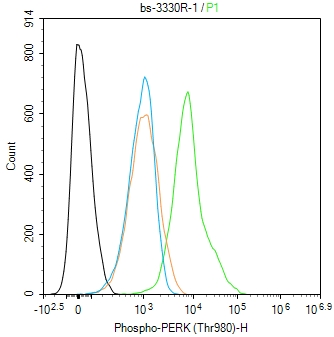
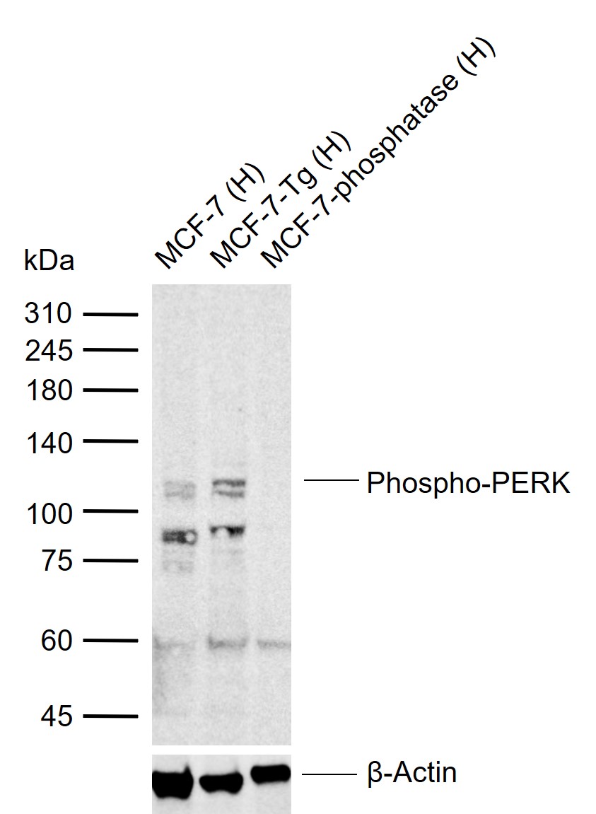
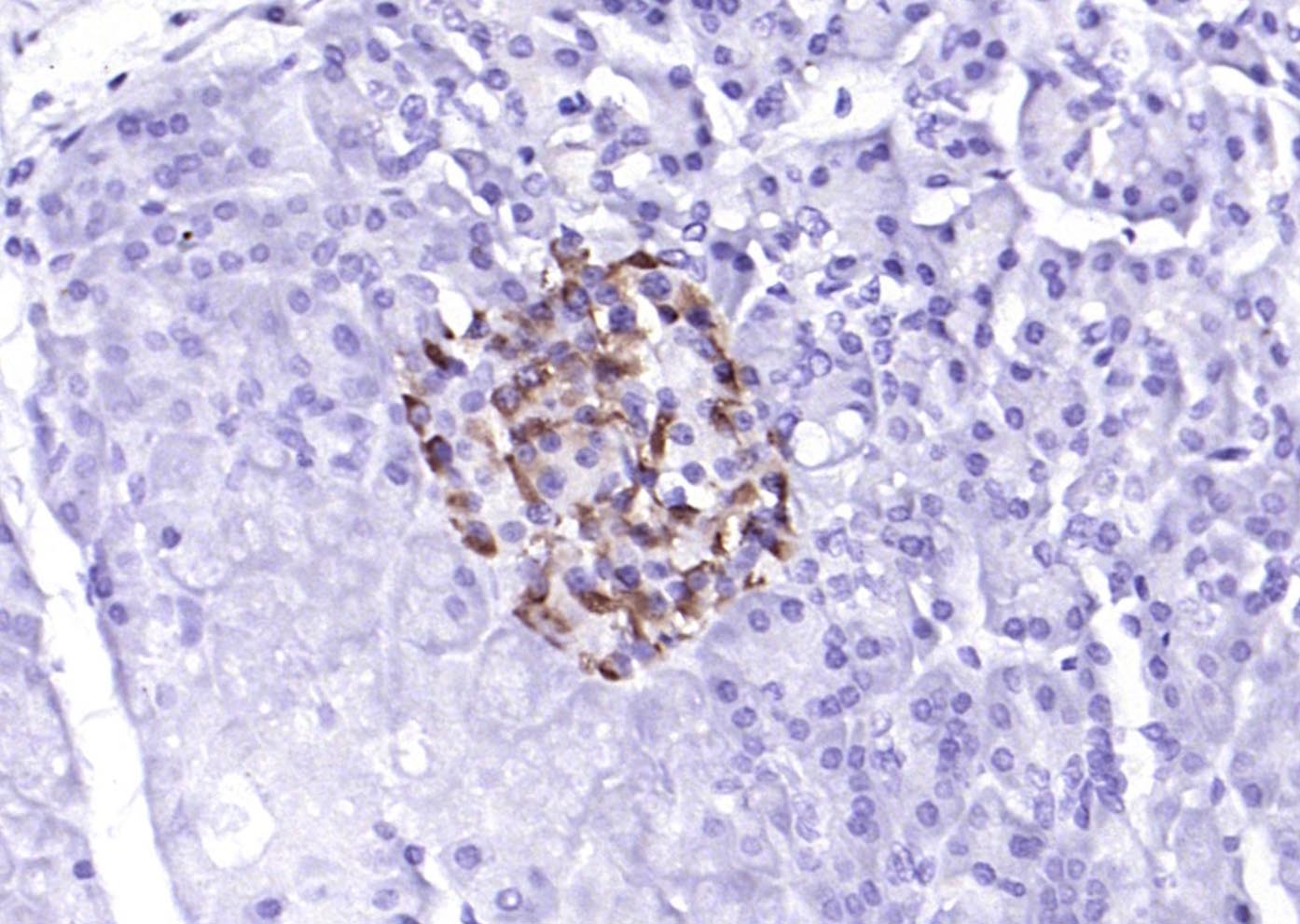
- 产品详情
- 实验流程
- 背景知识
Application 
| WB, IHC-P, IHC-F, IF |
|---|---|
| Primary Accession | Q9Z2B5 |
| Reactivity | Rat, Human, Mouse |
| Predicted | Pig, Dog, Rabbit |
| Host | Rabbit |
| Clonality | Polyclonal |
| Calculated MW | 124682 Da |
| Physical State | Liquid |
| Immunogen | KLH conjugated synthesised phosphopeptide derived from mouse PERK around the phosphorylation site of Thr980 |
| Epitope Specificity | H(p-T)GQ |
| Isotype | IgG |
| Purity | affinity purified by Protein A |
| Buffer | 0.01M TBS (pH7.4) with 1% BSA, 0.02% Proclin300 and 50% Glycerol. |
| SUBCELLULAR LOCATION | Endoplasmic reticulum membrane; Single-pass type I membrane protein. |
| DISEASE | Wolcott-Rallison syndrome (WRS) [MIM:226980]: A rare autosomal recessive disorder, characterized by permanent neonatal or early infancy insulin-dependent diabetes and, at a later age, epiphyseal dysplasia, osteoporosis, growth retardation and other multisystem manifestations, such as hepatic and renal dysfunctions, mental retardation and cardiovascular abnormalities. Note=The disease is caused by mutations affecting the gene represented in this entry. |
| Important Note | This product as supplied is intended for research use only, not for use in human, therapeutic or diagnostic applications. |
| Background Descriptions | The protein encoded by this gene phosphorylates the alpha subunit of eukaryotic translation-initiation factor 2 (EIF2), leading to its inactivation, and thus to a rapid reduction of translational initiation and repression of global protein synthesis. It is a type I membrane protein located in the endoplasmic reticulum (ER), where it is induced by ER stress caused by malfolded proteins. Mutations in this gene are associated with Wolcott-Rallison syndrome. [provided by RefSeq, Jan 2010]. |
| Gene ID | 13666 |
|---|---|
| Other Names | Eukaryotic translation initiation factor 2-alpha kinase 3, 2.7.11.1, PRKR-like endoplasmic reticulum kinase, Pancreatic eIF2-alpha kinase, 2.7.10.2, Eif2ak3 {ECO:0000312|MGI:MGI:1341830} |
| Dilution | WB=1:500-2000,IHC-P=1:100-500,IHC-F=1:100-500,IF=1:100-500,Flow-Cyt=2ug/Test |
| Storage | Store at -20 °C for one year. Avoid repeated freeze/thaw cycles. When reconstituted in sterile pH 7.4 0.01M PBS or diluent of antibody the antibody is stable for at least two weeks at 2-4 °C. |
For Research Use Only. Not For Use In Diagnostic Procedures.
| Name | Eif2ak3 {ECO:0000312|MGI:MGI:1341830} |
|---|---|
| Function | Metabolic-stress sensing protein kinase that phosphorylates the alpha subunit of eukaryotic translation initiation factor 2 (EIF2S1/eIF-2-alpha) in response to various stress, such as unfolded protein response (UPR) (PubMed:10882126, PubMed:11106749, PubMed:11430819, PubMed:11997520, PubMed:14978030, PubMed:16418533, PubMed:17998206, PubMed:21954288, PubMed:23921556, PubMed:9930704). Key effector of the integrated stress response (ISR) to unfolded proteins: EIF2AK3/PERK specifically recognizes and binds misfolded proteins, leading to its activation and EIF2S1/eIF-2-alpha phosphorylation (PubMed:11106749, PubMed:16418533, PubMed:17998206, PubMed:21954288, PubMed:23921556, PubMed:29386355, PubMed:9930704). EIF2S1/eIF-2-alpha phosphorylation in response to stress converts EIF2S1/eIF-2-alpha in a global protein synthesis inhibitor, leading to a global attenuation of cap-dependent translation, while concomitantly initiating the preferential translation of ISR-specific mRNAs, such as the transcriptional activators ATF4 and QRICH1, and hence allowing ATF4- and QRICH1-mediated reprogramming (PubMed:10882126, PubMed:11106749, PubMed:11430819, PubMed:11997520, PubMed:23921556). The EIF2AK3/PERK-mediated unfolded protein response increases mitochondrial oxidative phosphorylation by promoting ATF4-mediated expression of COX7A2L/SCAF1, thereby increasing formation of respiratory chain supercomplexes (PubMed:31023583). In contrast to most subcellular compartments, mitochondria are protected from the EIF2AK3/PERK-mediated unfolded protein response due to EIF2AK3/PERK inhibition by ATAD3A at mitochondria-endoplasmic reticulum contact sites (By similarity). In addition to EIF2S1/eIF-2-alpha, also phosphorylates NFE2L2/NRF2 in response to stress, promoting release of NFE2L2/NRF2 from the BCR(KEAP1) complex, leading to nuclear accumulation and activation of NFE2L2/NRF2 (PubMed:14517290, PubMed:14978030). Serves as a critical effector of unfolded protein response (UPR)-induced G1 growth arrest due to the loss of cyclin-D1 (CCND1) (PubMed:11035797). Involved in control of mitochondrial morphology and function (PubMed:23921556). |
| Cellular Location | Endoplasmic reticulum membrane; Single-pass type I membrane protein. Note=Localizes to the Localizes to endoplasmic reticulum membrane (PubMed:11430819). Also present at mitochondria-endoplasmic reticulum contact sites; where it interacts with ATAD3A (By similarity). {ECO:0000250|UniProtKB:Q9NZJ5, ECO:0000269|PubMed:11430819} |
| Tissue Location | Ubiquitous (PubMed:9930704). Highly expressed in mouse pancreas, an organ active in protein secretion (at protein level) (PubMed:11430819). |
Provided below are standard protocols that you may find useful for product applications.
BACKGROUND
This product as supplied is intended for research use only, not for use in human, therapeutic or diagnostic applications.
REFERENCES
Harding H.P.,et al.Nature 397:271-274(1999).
Harding H.P.,et al.Nature 398:90-90(1999).
Carninci P.,et al.Science 309:1559-1563(2005).
Bertolotti A.,et al.Nat. Cell Biol. 2:326-332(2000).
Brewer J.W.,et al.Proc. Natl. Acad. Sci. U.S.A. 97:12625-12630(2000).

终于等到您。ABCEPTA(百远生物)抗体产品。
点击下方“我要评价 ”按钮提交您的反馈信息,您的反馈和评价是我们最宝贵的财富之一,
我们将在1-3个工作日内处理您的反馈信息。
如有疑问,联系:0512-88856768 tech-china@abcepta.com.


