


 癌症的基本特征包括细胞增殖、血管生成、迁移、凋亡逃避机制和细胞永生等。找到癌症发生过程中这些通路的关键标记物和对应的抗体用于检测至关重要。
 为您推荐一个泛素化位点预测神器——泛素化分析工具,可以为您的蛋白的泛素化位点作出预测和评分。
 细胞自噬受体图形绘图工具为你的蛋白的细胞受体结合位点作出预测和评分,识别结合到自噬通路中的蛋白是非常重要的,便于让我们理解自噬在正常生理、病理过程中的作用,如发育、细胞分化、神经退化性疾病、压力条件下、感染和癌症。
Fyn Antibody
Purified Mouse Monoclonal Antibody (Mab)
- 产品详情
- 实验流程
- 背景知识
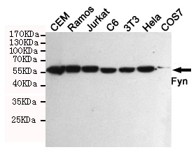
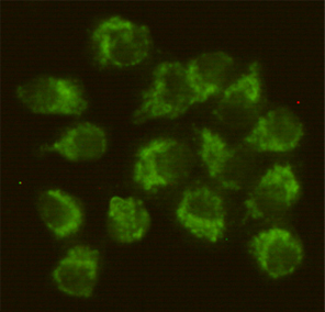
Application
| WB, ICC |
|---|---|
| Primary Accession | P06241 |
| Reactivity | Human, Mouse |
| Host | Mouse |
| Clonality | Monoclonal |
| Isotype | IgG2b |
| Calculated MW | 60762 Da |
| Gene ID | 2534 |
|---|---|
| Other Names | C syn protooncogene;Fyn;FYN oncogene related to SRC FGR YES;FYN_HUMAN;MGC45350;OKT3 induced calcium influx regulator;P59 FYN;p59-Fyn;Protein tyrosine kinase fyn;Proto oncogene tyrosine protein kinase fyn;Proto-oncogene c-Fyn;Proto-oncogene Syn;Protooncogene Syn;SLK;Src like kinase;Src yes related novel gene;Src-like kinase;Src/yes related novel;Src/yes related novel gene;SYN;Tyrosine kinase p59fyn T;Tyrosine kinase p59fyn(T);Tyrosine-protein kinase Fyn. |
| Dilution | WB~~1:500 ICC~~1:50 |
| Format | Liquid in PBS containing 50% glycerol, 0.5% BSA and 0.02% sodium azide, pH 7.3. |
| Storage | Store at 4°C short term. Aliquot and store at -20°C long term. Avoid freeze/thaw cycles. |
| Name | FYN |
|---|---|
| Function | Non-receptor tyrosine-protein kinase that plays a role in many biological processes including regulation of cell growth and survival, cell adhesion, integrin-mediated signaling, cytoskeletal remodeling, cell motility, immune response and axon guidance (PubMed:11536198, PubMed:15489916, PubMed:15557120, PubMed:16387660, PubMed:20100835, PubMed:7568038, PubMed:7822789). Inactive FYN is phosphorylated on its C-terminal tail within the catalytic domain (PubMed:15489916). Following activation by PKA, the protein subsequently associates with PTK2/FAK1, allowing PTK2/FAK1 phosphorylation, activation and targeting to focal adhesions (PubMed:15489916). Involved in the regulation of cell adhesion and motility through phosphorylation of CTNNB1 (beta-catenin) and CTNND1 (delta-catenin) (PubMed:17194753). Regulates cytoskeletal remodeling by phosphorylating several proteins including the actin regulator WAS and the microtubule-associated proteins MAP2 and MAPT (PubMed:14707117, PubMed:15536091). Promotes cell survival by phosphorylating AGAP2/PIKE- A and preventing its apoptotic cleavage (PubMed:16841086). Participates in signal transduction pathways that regulate the integrity of the glomerular slit diaphragm (an essential part of the glomerular filter of the kidney) by phosphorylating several slit diaphragm components including NPHS1, KIRREL1 and TRPC6 (PubMed:14761972, PubMed:18258597, PubMed:19179337). Plays a role in neural processes by phosphorylating DPYSL2, a multifunctional adapter protein within the central nervous system, ARHGAP32, a regulator for Rho family GTPases implicated in various neural functions, and SNCA, a small pre-synaptic protein (PubMed:11162638, PubMed:12788081, PubMed:19652227). Involved in reelin signaling by mediating phosphorylation of DAB1 following reelin (RELN)- binding to its receptor (By similarity). Participates in the downstream signaling pathways that lead to T-cell differentiation and proliferation following T-cell receptor (TCR) stimulation (PubMed:22080863). Phosphorylates PTK2B/PYK2 in response to T-cell receptor activation (PubMed:20028775). Also participates in negative feedback regulation of TCR signaling through phosphorylation of PAG1 and PDCD1 (PubMed:18056706, PubMed:32184441). Phosphorylation of PAG1 promotes interaction between PAG1 and CSK and recruitment of CSK to lipid rafts (PubMed:18056706). Phosphorylation of PDCD1 leads to the recruitment of PTPN11/SHP-2 that mediates dephosphorylation of key TCR proximal signaling molecules (PubMed:32184441). CSK maintains LCK and FYN in an inactive form (By similarity). Promotes CD28-induced phosphorylation of VAV1 (PubMed:11005864). In mast cells, phosphorylates CLNK after activation of immunoglobulin epsilon receptor signaling (By similarity). Can also promote CD244-mediated NK cell activation (PubMed:15713798). |
| Cellular Location | Cytoplasm. Nucleus Cell membrane. Perikaryon {ECO:0000250|UniProtKB:Q62844} Note=Present and active in lipid rafts (PubMed:12218089) Palmitoylation is crucial for proper trafficking (PubMed:8206991) |
| Tissue Location | Isoform 1 is highly expressed in the brain. Isoform 2 is expressed in cells of hemopoietic lineages, especially T- lymphocytes. |
For Research Use Only. Not For Use In Diagnostic Procedures.
Provided below are standard protocols that you may find useful for product applications.
BACKGROUND
Non-receptor tyrosine-protein kinase that plays a role in many biological processes including regulation of cell growth and survival, cell adhesion, integrin-mediated signaling, cytoskeletal remodeling, cell motility, immune response and axon guidance. Inactive FYN is phosphorylated on its C-terminal tail within the catalytic domain. Following activation by PKA, the protein subsequently associates with PTK2/FAK1, allowing PTK2/FAK1 phosphorylation, activation and targeting to focal adhesions. Involved in the regulation of cell adhesion and motility through phosphorylation of CTNNB1 (beta-catenin) and CTNND1 (delta- catenin). Regulates cytoskeletal remodeling by phosphorylating several proteins including the actin regulator WAS and the microtubule-associated proteins MAP2 and MAPT. Promotes cell survival by phosphorylating AGAP2/PIKE-A and preventing its apoptotic cleavage. Participates in signal transduction pathways that regulate the integrity of the glomerular slit diaphragm (an essential part of the glomerular filter of the kidney) by phosphorylating several slit diaphragm components including NPHS1, KIRREL and TRPC6. Plays a role in neural processes by phosphorylating DPYSL2, a multifunctional adapter protein within the central nervous system, ARHGAP32, a regulator for Rho family GTPases implicated in various neural functions, and SNCA, a small pre-synaptic protein. Participates in the downstream signaling pathways that lead to T-cell differentiation and proliferation following T-cell receptor (TCR) stimulation. Also participates in negative feedback regulation of TCR signaling through phosphorylation of PAG1, thereby promoting interaction between PAG1 and CSK and recruitment of CSK to lipid rafts. CSK maintains LCK and FYN in an inactive form. Promotes CD28-induced phosphorylation of VAV1.
REFERENCES
Kawakami T.,et al.Mol. Cell. Biol. 6:4195-4201(1986).
Semba K.,et al.Proc. Natl. Acad. Sci. U.S.A. 83:5459-5463(1986).
Rigley K.,et al.J. Immunol. 154:1136-1145(1995).
Goshima N.,et al.Nat. Methods 5:1011-1017(2008).
Mungall A.J.,et al.Nature 425:805-811(2003).
终于等到您。ABCEPTA(百远生物)抗体产品。
点击下方“我要评价 ”按钮提交您的反馈信息,您的反馈和评价是我们最宝贵的财富之一,
我们将在1-3个工作日内处理您的反馈信息。
如有疑问,联系:0512-88856768 tech-china@abcepta.com.


