


 癌症的基本特征包括细胞增殖、血管生成、迁移、凋亡逃避机制和细胞永生等。找到癌症发生过程中这些通路的关键标记物和对应的抗体用于检测至关重要。
 为您推荐一个泛素化位点预测神器——泛素化分析工具,可以为您的蛋白的泛素化位点作出预测和评分。
 细胞自噬受体图形绘图工具为你的蛋白的细胞受体结合位点作出预测和评分,识别结合到自噬通路中的蛋白是非常重要的,便于让我们理解自噬在正常生理、病理过程中的作用,如发育、细胞分化、神经退化性疾病、压力条件下、感染和癌症。
CD248 Antibody
Purified Rabbit Polyclonal Antibody (Pab)
- 产品详情
- 实验流程
- 背景知识
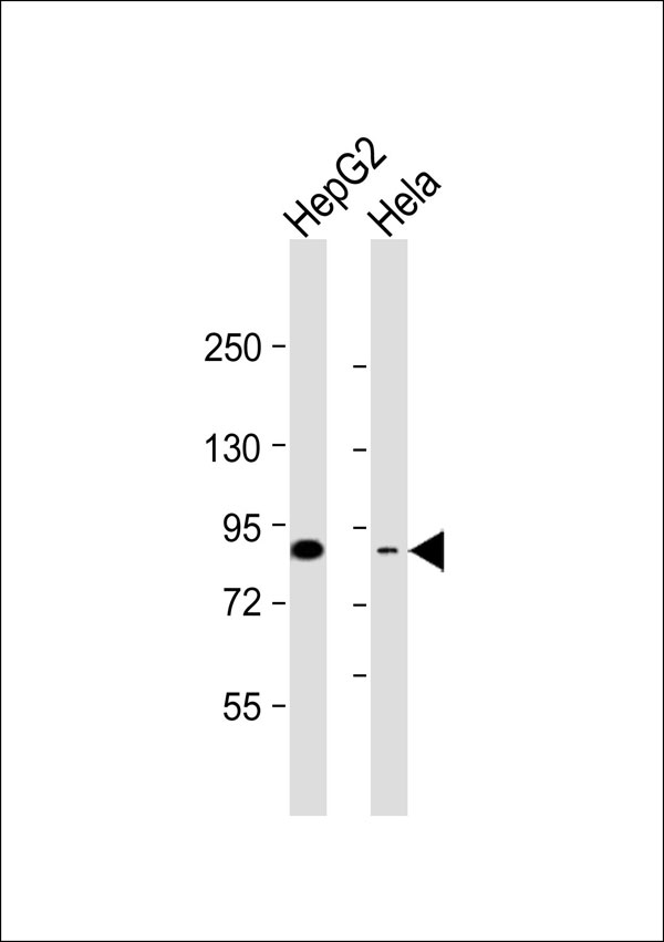
Application
| WB |
|---|---|
| Primary Accession | Q9HCU0 |
| Reactivity | Human |
| Host | Rabbit |
| Clonality | Polyclonal |
| Calculated MW | 80859 Da |
| Gene ID | 57124 |
|---|---|
| Other Names | Endosialin, Tumor endothelial marker 1, CD248, CD248, CD164L1, TEM1 |
| Target/Specificity | KLH-conjugated synthetic peptide encompassing a sequence within the center region of human CD248. The exact sequence is proprietary. |
| Dilution | WB~~ 1:1000 |
| Format | Rabbit IgG in phosphate buffered saline , pH 7.4, 150mM NaCl, 0.09% (W/V) sodium azide and 50% glycerol |
| Storage | Store at -20 °C.Stable for 12 months from date of receipt |
| Name | CD248 |
|---|---|
| Synonyms | CD164L1, TEM1 |
| Function | Cell surface glycoprotein involved in various biological processes including angiogenesis, immune response modulation, and tissue remodeling and repair. Participates in pericyte proliferation through positive modulation of the PDGF receptor signaling pathway (PubMed:20484976). Acts as a scaffold for factor X, triggering allosteric changes and the spatial re-alignment of factor X with the TF-factor VIIa complex, thereby enhancing coagulation activation. Modulates the insulin signaling pathway by interacting with insulin receptor/INSR and by diminishing its capacity to be autophosphorylated in response to insulin. Also regulates LPS-induced inflammatory response in macrophages by favoring the production of proinflammatory cytokines. In human, negatively regulates T-cell proliferation compared with stromal cells where it increases proliferation (PubMed:21466550). |
| Cellular Location | Membrane; Single-pass type I membrane protein |
| Tissue Location | Expressed in tumor endothelial cells but absent or barely detectable in normal endothelial cells. Expressed in metastatic lesions of the liver and during angiogenesis of corpus luteum formation and wound healing. Expressed in vascular endothelial cells of malignant tumors but not in normal blood vessels. Expressed in stromal fibroblasts. Strongly expressed in pericytes (PubMed:20484976) Expressed on stromal cells and cells with lymphoid morphology such a T- cells (PubMed:21466550). |
Research Areas
For Research Use Only. Not For Use In Diagnostic Procedures.
Application Protocols
Provided below are standard protocols that you may find useful for product applications.
BACKGROUND
May play a role in tumor angiogenesis.
REFERENCES
St Croix B.,et al.Science 289:1197-1202(2000).
Christian S.,et al.J. Biol. Chem. 276:7408-7414(2001).
Ota T.,et al.Nat. Genet. 36:40-45(2004).
Rettig W.J.,et al.Proc. Natl. Acad. Sci. U.S.A. 89:10832-10836(1992).
Dolznig H.,et al.Cancer Immun. 5:10-10(2005).
终于等到您。ABCEPTA(百远生物)抗体产品。
点击下方“我要评价 ”按钮提交您的反馈信息,您的反馈和评价是我们最宝贵的财富之一,
我们将在1-3个工作日内处理您的反馈信息。
如有疑问,联系:0512-88856768 tech-china@abcepta.com.
¥ 1,500.00
Cat# AP53290


