


 癌症的基本特征包括细胞增殖、血管生成、迁移、凋亡逃避机制和细胞永生等。找到癌症发生过程中这些通路的关键标记物和对应的抗体用于检测至关重要。
 为您推荐一个泛素化位点预测神器——泛素化分析工具,可以为您的蛋白的泛素化位点作出预测和评分。
 细胞自噬受体图形绘图工具为你的蛋白的细胞受体结合位点作出预测和评分,识别结合到自噬通路中的蛋白是非常重要的,便于让我们理解自噬在正常生理、病理过程中的作用,如发育、细胞分化、神经退化性疾病、压力条件下、感染和癌症。
DLAT Antibody (C-term)
Affinity Purified Rabbit Polyclonal Antibody (Pab)
- 产品详情
- 实验流程
- 背景知识
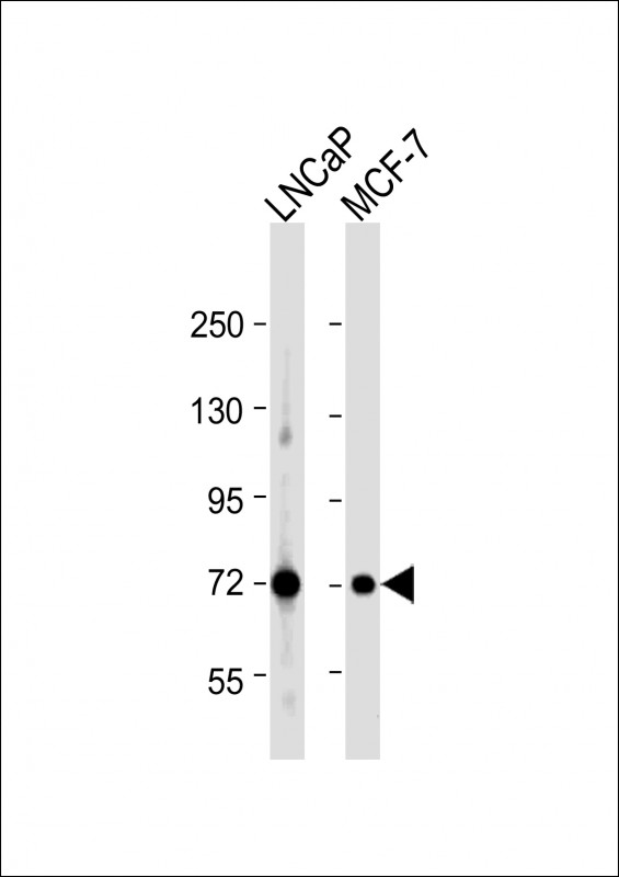
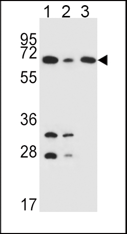
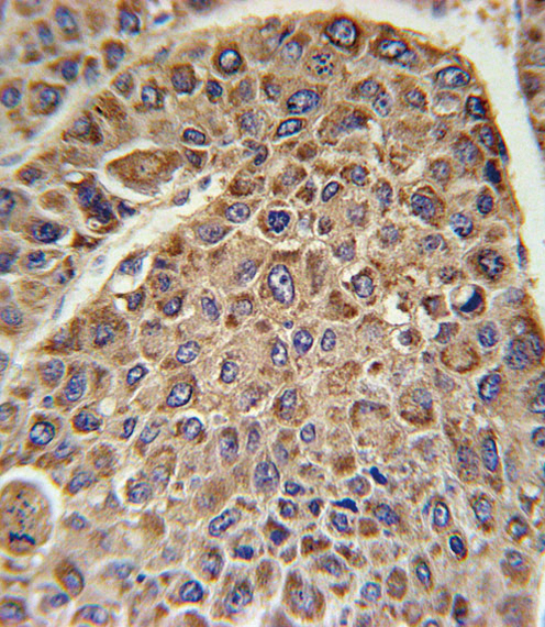
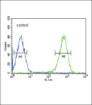
Application
| WB, IHC-P, FC, E |
|---|---|
| Primary Accession | P10515 |
| Other Accession | NP_001922.2 |
| Reactivity | Human |
| Host | Rabbit |
| Clonality | Polyclonal |
| Isotype | Rabbit IgG |
| Calculated MW | 68997 Da |
| Antigen Region | 579-607 aa |
| Gene ID | 1737 |
|---|---|
| Other Names | Dihydrolipoyllysine-residue acetyltransferase component of pyruvate dehydrogenase complex, mitochondrial, 70 kDa mitochondrial autoantigen of primary biliary cirrhosis, PBC, Dihydrolipoamide acetyltransferase component of pyruvate dehydrogenase complex, M2 antigen complex 70 kDa subunit, Pyruvate dehydrogenase complex component E2, PDC-E2, PDCE2, DLAT, DLTA |
| Target/Specificity | This DLAT antibody is generated from rabbits immunized with a KLH conjugated synthetic peptide between 579-607 amino acids from the C-terminal region of human DLAT. |
| Dilution | WB~~1:1000 IHC-P~~1:100~500 FC~~1:10~50 E~~Use at an assay dependent concentration. |
| Format | Purified polyclonal antibody supplied in PBS with 0.09% (W/V) sodium azide. This antibody is purified through a protein A column, followed by peptide affinity purification. |
| Storage | Maintain refrigerated at 2-8°C for up to 2 weeks. For long term storage store at -20°C in small aliquots to prevent freeze-thaw cycles. |
| Precautions | DLAT Antibody (C-term) is for research use only and not for use in diagnostic or therapeutic procedures. |
| Name | DLAT (HGNC:2896) |
|---|---|
| Synonyms | DLTA |
| Function | The pyruvate dehydrogenase (PDH) complex, catalyzes the overall conversion of pyruvate to acetyl-CoA and CO(2), and thereby links cytoplasmic glycolysis and the mitochondrial tricarboxylic acid (TCA) cycle (Probable). It contains multiple copies of three enzymatic components: pyruvate dehydrogenase (E1), dihydrolipoamide acetyltransferase (E2) and dihydrolipoamide dehydrogenase (E3); (Probable). Within this complex, the catalytic function of this enzyme is to accept, and to transfer to coenzyme A, acetyl groups from acetyl- lipoyl moiety generated by the pyruvate dehydrogenase, leading to acetyl-CoA formation (Probable). |
| Cellular Location | Mitochondrion matrix {ECO:0000250|UniProtKB:P08461} |
For Research Use Only. Not For Use In Diagnostic Procedures.
Provided below are standard protocols that you may find useful for product applications.
BACKGROUND
DLAT encodes component E2 of the multi-enzyme pyruvate dehydrogenase complex (PDC). PDC resides in the inner mitochondrial membrane and catalyzes the conversion of pyruvate to acetyl coenzyme A. The protein product of this gene, dihydrolipoamide acetyltransferase, accepts acetyl groups formed by the oxidative decarboxylation of pyruvate and transfers them to coenzyme A. Dihydrolipoamide acetyltransferase is the antigen for antimitochondrial antibodies. These autoantibodies are present in nearly 95% of patients with the autoimmune liver disease primary biliary cirrhosis (PBC). In PBC, activated T lymphocytes attack and destroy epithelial cells in the bile duct where this protein is abnormally distributed and overexpressed. PBC enventually leads to cirrhosis and liver failure.
REFERENCES
Trynka, G., et al. Gut 58(8):1078-1083(2009)
Lleo, A., et al. Hepatology 49(3):871-879(2009)
Korotchkina, L.G., et al. FEBS Lett. 582(3):468-472(2008)
终于等到您。ABCEPTA(百远生物)抗体产品。
点击下方“我要评价 ”按钮提交您的反馈信息,您的反馈和评价是我们最宝贵的财富之一,
我们将在1-3个工作日内处理您的反馈信息。
如有疑问,联系:0512-88856768 tech-china@abcepta.com.


