


 癌症的基本特征包括细胞增殖、血管生成、迁移、凋亡逃避机制和细胞永生等。找到癌症发生过程中这些通路的关键标记物和对应的抗体用于检测至关重要。
 为您推荐一个泛素化位点预测神器——泛素化分析工具,可以为您的蛋白的泛素化位点作出预测和评分。
 细胞自噬受体图形绘图工具为你的蛋白的细胞受体结合位点作出预测和评分,识别结合到自噬通路中的蛋白是非常重要的,便于让我们理解自噬在正常生理、病理过程中的作用,如发育、细胞分化、神经退化性疾病、压力条件下、感染和癌症。
Anti-MARK Antibody
- 产品详情
- 实验流程
- 背景知识
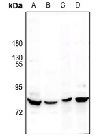
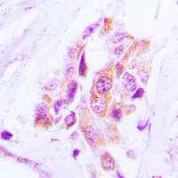
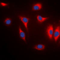
Application
| WB, IF |
|---|---|
| Primary Accession | Q9P0L2 |
| Other Accession | Q7KZI7, P27448, Q96L34 |
| Reactivity | Human, Mouse, Rat |
| Host | Rabbit |
| Clonality | Polyclonal |
| Calculated MW | 89003 Da |
| Gene ID | 4139 |
|---|---|
| Other Names | MARK1; KIAA1477; MARK; Serine/threonine-protein kinase MARK1; MAP/microtubule affinity-regulating kinase 1; PAR1 homolog c; Par-1c; Par1c; MARK2; EMK1; Serine/threonine-protein kinase MARK2; ELKL motif kinase 1; EMK-1; MAP/microtubule affinity-regulating kinase 2; PAR1 homolog; PAR1 homolog b; Par-1b; Par1b; MARK3; CTAK1; EMK2; MAP/microtubule affinity-regulating kinase 3; C-TAK1; cTAK1; Cdc25C-associated protein kinase 1; ELKL motif kinase 2; EMK-2; Protein kinase STK10; Ser/Thr protein kinase PAR-1; Par-1a; Serine/threonine-protein kinase p78; MARK4; KIAA1860; MARKL1; MAP/microtubule affinity-regulating kinase 4; MAP/microtubule affinity-regulating kinase-like 1 |
| Target/Specificity | KLH-conjugated synthetic peptide encompassing a sequence within the center region of human MARK. The exact sequence is proprietary. |
| Dilution | WB~~1/500 - 1/1000 IF~~1/50 - 1/200 |
| Format | Liquid in 0.42% Potassium phosphate, 0.87% Sodium chloride, pH 7.3, 30% glycerol, and 0.09% (W/V) sodium azide. |
| Storage | Store at -20 °C.Stable for 12 months from date of receipt |
| Name | MARK1 (HGNC:6896) |
|---|---|
| Function | Serine/threonine-protein kinase (PubMed:23666762). Involved in cell polarity and microtubule dynamics regulation. Phosphorylates DCX, MAP2 and MAP4. Phosphorylates the microtubule-associated protein MAPT/TAU (PubMed:23666762). Involved in cell polarity by phosphorylating the microtubule-associated proteins MAP2, MAP4 and MAPT/TAU at KXGS motifs, causing detachment from microtubules, and their disassembly. Involved in the regulation of neuronal migration through its dual activities in regulating cellular polarity and microtubule dynamics, possibly by phosphorylating and regulating DCX. Also acts as a positive regulator of the Wnt signaling pathway, probably by mediating phosphorylation of dishevelled proteins (DVL1, DVL2 and/or DVL3). |
| Cellular Location | Cell membrane; Peripheral membrane protein. Cytoplasm, cytoskeleton. Cytoplasm Cell projection, dendrite. Note=Appears to localize to an intracellular network. |
| Tissue Location | Highly expressed in heart, skeletal muscle, brain, fetal brain and fetal kidney. |
Research Areas
For Research Use Only. Not For Use In Diagnostic Procedures.
Application Protocols
Provided below are standard protocols that you may find useful for product applications.
BACKGROUND
Rabbit polyclonal antibody to MARK
终于等到您。ABCEPTA(百远生物)抗体产品。
点击下方“我要评价 ”按钮提交您的反馈信息,您的反馈和评价是我们最宝贵的财富之一,
我们将在1-3个工作日内处理您的反馈信息。
如有疑问,联系:0512-88856768 tech-china@abcepta.com.
¥ 1,500.00
Cat# AP53905


