


 癌症的基本特征包括细胞增殖、血管生成、迁移、凋亡逃避机制和细胞永生等。找到癌症发生过程中这些通路的关键标记物和对应的抗体用于检测至关重要。
 为您推荐一个泛素化位点预测神器——泛素化分析工具,可以为您的蛋白的泛素化位点作出预测和评分。
 细胞自噬受体图形绘图工具为你的蛋白的细胞受体结合位点作出预测和评分,识别结合到自噬通路中的蛋白是非常重要的,便于让我们理解自噬在正常生理、病理过程中的作用,如发育、细胞分化、神经退化性疾病、压力条件下、感染和癌症。
Anti-Tensin 3 Antibody
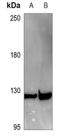
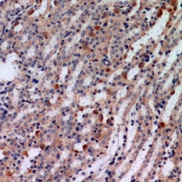
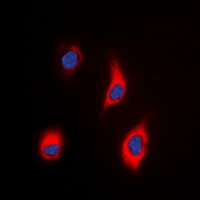
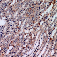
- 产品详情
- 实验流程
Application 
| WB, IHC, IF/IC |
|---|---|
| Primary Accession | Q68CZ2 |
| Reactivity | Human, Rat |
| Host | Rabbit |
| Clonality | Polyclonal |
| Calculated MW | 155266 Da |
| Gene ID | 64759 |
|---|---|
| Other Names | TEM6; TENS1; TPP; Tensin-3; Tensin-like SH2 domain-containing protein 1; Tumor endothelial marker 6 |
| Target/Specificity | KLH-conjugated synthetic peptide encompassing a sequence within the center region of human Tensin 3. The exact sequence is proprietary. |
| Dilution | WB~~WB (1/500 - 1/2000) IHC~~1/50 - 1/200 IF/IC~~N/A |
| Format | Liquid in 0.42% Potassium phosphate, 0.87% Sodium chloride, pH 7.3, 30% glycerol, and 0.01% sodium azide. |
| Storage | Store at -20 °C.Stable for 12 months from date of receipt |
For Research Use Only. Not For Use In Diagnostic Procedures.
| Name | TNS3 |
|---|---|
| Synonyms | TEM6, TENS1, TPP |
| Function | May act as a protein phosphatase and/or a lipid phosphatase (Probable). Involved in the dissociation of the integrin-tensin-actin complex (PubMed:17643115). EGF activates TNS4 and down-regulates TNS3 which results in capping the tail of ITGB1 (PubMed:17643115). Increases DOCK5 guanine nucleotide exchange activity towards Rac and plays a role in osteoclast podosome organization (By similarity). Enhances RHOA activation in the presence of DLC1 (PubMed:26427649). Required for growth factor-induced epithelial cell migration; growth factor stimulation induces TNS3 phosphorylation which changes its binding preference from DLC1 to the p85 regulatory subunit of the PI3K kinase complex, displacing PI3K inhibitor PTEN and resulting in translocation of the TNS3-p85 complex to the leading edge of migrating cells to promote RAC1 activation (PubMed:26166433). Meanwhile, PTEN switches binding preference from p85 to DLC1 and the PTEN-DLC1 complex translocates to the posterior of migrating cells to activate RHOA (PubMed:26166433). Acts as an adapter protein by bridging the association of scaffolding protein PEAK1 with integrins ITGB1, ITGB3 and ITGB5 which contributes to the promotion of cell migration (PubMed:35687021). Controls tonsil-derived mesenchymal stem cell proliferation and differentiation by regulating the activity of integrin ITGB1 (PubMed:31905841). |
| Cellular Location | Cell junction, focal adhesion. Cell projection, podosome {ECO:0000250|UniProtKB:Q5SSZ5}. Note=Localizes to both focal and fibrillar adhesions but is mostly found in fibrillar adhesions. |
| Tissue Location | Expressed in umbilical vein endothelial cells, epithelial cells, and fibroblasts cells (at protein level). Highly expressed in thyroid, kidney and placenta. Low expression in heart, skeletal muscle, spleen, liver, and lung. Expressed at higher levels in tonsil-derived mesenchymal stem cells (MSCs) than in adipose tissue- derived MSCs or bone marrow-derived MSCs (PubMed:31905841). Expressed in tumor endothelial cells. Expression seems to be down-regulated in thyroid tumor tissues and in anaplastic carcinomas |
Research Areas
Application Protocols
Provided below are standard protocols that you may find useful for product applications.

终于等到您。ABCEPTA(百远生物)抗体产品。
点击下方“我要评价 ”按钮提交您的反馈信息,您的反馈和评价是我们最宝贵的财富之一,
我们将在1-3个工作日内处理您的反馈信息。
如有疑问,联系:0512-88856768 tech-china@abcepta.com.


