


 癌症的基本特征包括细胞增殖、血管生成、迁移、凋亡逃避机制和细胞永生等。找到癌症发生过程中这些通路的关键标记物和对应的抗体用于检测至关重要。
 为您推荐一个泛素化位点预测神器——泛素化分析工具,可以为您的蛋白的泛素化位点作出预测和评分。
 细胞自噬受体图形绘图工具为你的蛋白的细胞受体结合位点作出预测和评分,识别结合到自噬通路中的蛋白是非常重要的,便于让我们理解自噬在正常生理、病理过程中的作用,如发育、细胞分化、神经退化性疾病、压力条件下、感染和癌症。
Anti-CD66a/b/c Antibody
- 产品详情
- 实验流程
- 背景知识
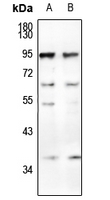
Application
| WB |
|---|---|
| Primary Accession | P13688 |
| Other Accession | P40199, P31997 |
| Reactivity | Human, Mouse |
| Host | Rabbit |
| Clonality | Polyclonal |
| Calculated MW | 57560 Da |
| Gene ID | 634 |
|---|---|
| Other Names | CEACAM1; BGP; BGP1; Carcinoembryonic antigen-related cell adhesion molecule 1; Biliary glycoprotein 1; BGP-1; CD66a; CEACAM6; NCA; Carcinoembryonic antigen-related cell adhesion molecule 6; Non-specific crossreacting antigen; Normal cross-reacting antigen; CD66c; CEACAM8; CGM6; Carcinoembryonic antigen-related cell adhesion molecule 8; CD67 antigen; Carcinoembryonic antigen CGM6; Non-specific cross-reacting antigen NCA-95; CD66b |
| Target/Specificity | KLH-conjugated synthetic peptide encompassing a sequence within the center region of human CD66a/b/c. The exact sequence is proprietary. |
| Dilution | WB~~1/500 - 1/1000 |
| Format | Liquid in 0.42% Potassium phosphate, 0.87% Sodium chloride, pH 7.3, 30% glycerol, and 0.09% (W/V) sodium azide. |
| Storage | Store at -20 °C.Stable for 12 months from date of receipt |
| Name | CEACAM1 (HGNC:1814) |
|---|---|
| Function | [Isoform 1]: Cell adhesion protein that mediates homophilic cell adhesion in a calcium-independent manner (By similarity). Plays a role as coinhibitory receptor in immune response, insulin action and also functions as an activator during angiogenesis (PubMed:18424730, PubMed:23696226, PubMed:25363763). Its coinhibitory receptor function is phosphorylation- and PTPN6 -dependent, which in turn, suppress signal transduction of associated receptors by dephosphorylation of their downstream effectors. Plays a role in immune response, of T cells, natural killer (NK) and neutrophils (PubMed:18424730, PubMed:23696226). Upon TCR/CD3 complex stimulation, inhibits TCR- mediated cytotoxicity by blocking granule exocytosis by mediating homophilic binding to adjacent cells, allowing interaction with and phosphorylation by LCK and interaction with the TCR/CD3 complex which recruits PTPN6 resulting in dephosphorylation of CD247 and ZAP70 (PubMed:18424730). Also inhibits T cell proliferation and cytokine production through inhibition of JNK cascade and plays a crucial role in regulating autoimmunity and anti-tumor immunity by inhibiting T cell through its interaction with HAVCR2 (PubMed:25363763). Upon natural killer (NK) cells activation, inhibit KLRK1-mediated cytolysis of CEACAM1-bearing tumor cells by trans-homophilic interactions with CEACAM1 on the target cell and lead to cis-interaction between CEACAM1 and KLRK1, allowing PTPN6 recruitment and then VAV1 dephosphorylation (PubMed:23696226). Upon neutrophils activation negatively regulates IL1B production by recruiting PTPN6 to a SYK-TLR4-CEACAM1 complex, that dephosphorylates SYK, reducing the production of reactive oxygen species (ROS) and lysosome disruption, which in turn, reduces the activity of the inflammasome. Down-regulates neutrophil production by acting as a coinhibitory receptor for CSF3R by down-regulating the CSF3R-STAT3 pathway through recruitment of PTPN6 that dephosphorylates CSF3R (By similarity). Also regulates insulin action by promoting INS clearance and regulating lipogenesis in liver through regulating insulin signaling (By similarity). Upon INS stimulation, undergoes phosphorylation by INSR leading to INS clearance by increasing receptor-mediated insulin endocytosis. This inernalization promotes interaction with FASN leading to receptor-mediated insulin degradation and to reduction of FASN activity leading to negative regulation of fatty acid synthesis. INSR-mediated phosphorylation also provokes a down-regulation of cell proliferation through SHC1 interaction resulting in decrease coupling of SHC1 to the MAPK3/ERK1-MAPK1/ERK2 and phosphatidylinositol 3-kinase pathways (By similarity). Functions as activator in angiogenesis by promoting blood vessel remodeling through endothelial cell differentiation and migration and in arteriogenesis by increasing the number of collateral arteries and collateral vessel calibers after ischemia. Also regulates vascular permeability through the VEGFR2 signaling pathway resulting in control of nitric oxide production (By similarity). Down-regulates cell growth in response to EGF through its interaction with SHC1 that mediates interaction with EGFR resulting in decrease coupling of SHC1 to the MAPK3/ERK1- MAPK1/ERK2 pathway (By similarity). Negatively regulates platelet aggregation by decreasing platelet adhesion on type I collagen through the GPVI-FcRgamma complex (By similarity). Inhibits cell migration and cell scattering through interaction with FLNA; interferes with the interaction of FLNA with RALA (PubMed:16291724). Mediates bile acid transport activity in a phosphorylation dependent manner (By similarity). Negatively regulates osteoclastogenesis (By similarity). |
| Cellular Location | [Isoform 1]: Cell membrane {ECO:0000250|UniProtKB:P16573}; Single-pass type I membrane protein {ECO:0000250|UniProtKB:P16573}. Lateral cell membrane {ECO:0000250|UniProtKB:P16573}. Apical cell membrane {ECO:0000250|UniProtKB:P16573}. Basal cell membrane {ECO:0000250|UniProtKB:P16573}. Cell junction. Cell junction, adherens junction {ECO:0000250|UniProtKB:P16573}. Note=Canalicular domain of hepatocyte plasma membranes. Found as a mixture of monomer, dimer and oligomer in the plasma membrane. Occurs predominantly as cis-dimers and/or small cis-oligomers in the cell junction regions. Found as dimer in the solution. Predominantly localized to the lateral cell membranes {ECO:0000250|UniProtKB:P16573} [Isoform 3]: Secreted [Isoform 5]: Cell membrane; Single-pass type I membrane protein [Isoform 7]: Cell membrane; Single-pass type I membrane protein Cell projection, microvillus membrane {ECO:0000250|UniProtKB:P31809}; Single-pass type I membrane protein. Apical cell membrane; Single-pass type I membrane protein. Note=Localized to the apical glycocalyx surface (PubMed:10436421). Colocalizes with CEACAM20 at the apical brush border of intestinal cells {ECO:0000250|UniProtKB:P31809, ECO:0000269|PubMed:10436421} |
| Tissue Location | Expressed in columnar epithelial cells of the colon (at protein level) (PubMed:10436421). The predominant forms expressed by T cells are those containing a long cytoplasmic domain (PubMed:18424730). Expressed in granulocytes and lymphocytes Leukocytes only express isoforms 6 and isoform 1 (PubMed:11994468) |
Research Areas
For Research Use Only. Not For Use In Diagnostic Procedures.
Application Protocols
Provided below are standard protocols that you may find useful for product applications.
BACKGROUND
Rabbit polyclonal antibody to CD66a/b/c
终于等到您。ABCEPTA(百远生物)抗体产品。
点击下方“我要评价 ”按钮提交您的反馈信息,您的反馈和评价是我们最宝贵的财富之一,
我们将在1-3个工作日内处理您的反馈信息。
如有疑问,联系:0512-88856768 tech-china@abcepta.com.
¥ 1,500.00
Cat# AP54056


