


 癌症的基本特征包括细胞增殖、血管生成、迁移、凋亡逃避机制和细胞永生等。找到癌症发生过程中这些通路的关键标记物和对应的抗体用于检测至关重要。
 为您推荐一个泛素化位点预测神器——泛素化分析工具,可以为您的蛋白的泛素化位点作出预测和评分。
 细胞自噬受体图形绘图工具为你的蛋白的细胞受体结合位点作出预测和评分,识别结合到自噬通路中的蛋白是非常重要的,便于让我们理解自噬在正常生理、病理过程中的作用,如发育、细胞分化、神经退化性疾病、压力条件下、感染和癌症。
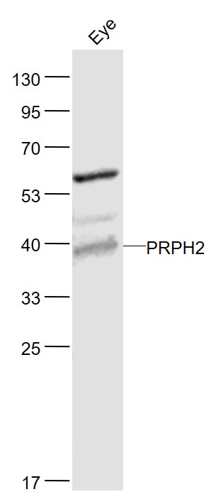
PRPH2 Rabbit pAb
PRPH2 Rabbit pAb
- 产品详情
- 实验流程
- 背景知识
Application
| WB |
|---|---|
| Primary Accession | P23942 |
| Reactivity | Mouse |
| Predicted | Human, Rat, Chicken, Dog, Horse, Sheep |
| Host | Rabbit |
| Clonality | Polyclonal |
| Calculated MW | 39272 Da |
| Physical State | Liquid |
| Immunogen | KLH conjugated synthetic peptide derived from human PRPH2 |
| Epitope Specificity | 131-230/346 |
| Isotype | IgG |
| Purity | affinity purified by Protein A |
| Buffer | 0.01M TBS (pH7.4) with 1% BSA, 0.02% Proclin300 and 50% Glycerol. |
| SUBCELLULAR LOCATION | Membrane; Multi-pass membrane protein. |
| SIMILARITY | Belongs to the PRPH2/ROM1 family. |
| SUBUNIT | Homodimer; disulfide-linked. Probably forms a complex with a ROM1 homodimer. Other proteins could associate with this complex in rods. Interacts with MREG. |
| DISEASE | Defects in PRPH2 are the cause of retinitis pigmentosa type 7 (RP7). RP leads to degeneration of retinal photoreceptor cells. Patients typically have night vision blindness and loss of midperipheral visual field. As their condition progresses, they lose their far peripheral visual field and eventually central vision as well. Defects in PRPH2 are a cause of retinitis punctata albescens. Defects in PRPH2 are a cause of adult-onset vitelliform macular dystrophy (AVMD). AVMD is a rare autosomal dominant disorder with incomplete penetrance and highly variable expression. Patients usually become symptomatic in the fourth or fifth decade of life with a protracted disease of decreased visual acuity. Defects in PRPH2 are a cause of patterned dystrophy of retinal pigment epithelium (PDREP). Patterned dystrophies of the retinal pigment epithelium (RPE) refer to a heterogeneous group of macular disorders. Three main types of PDREP have been described: reticular (fishnet-like) dystrophy, macroreticular (spider-shaped) dystrophy and butterfly-shaped pigment dystrophy. Defects in PRPH2 are a cause of choroidal dystrophy central areolar type 2 (CACD2). It is a disorder which affects the posterior pole of the eye, and early lesions consist of a non-specific area of granular hyperpigmentation at the fovea. The characteristic sign of the disorder, a zone of atrophy that develops in the macula of the eye and involves the retinal pigment epithelium and the choriocapillaris, occurs several decades after onset. |
| Important Note | This product as supplied is intended for research use only, not for use in human, therapeutic or diagnostic applications. |
| Background Descriptions | May function as an adhesion molecule involved in stabilization and compaction of outer segment disks or in the maintenance of the curvature of the rim. It is essential for disk morphogenesis. |
| Gene ID | 5961 |
|---|---|
| Other Names | Peripherin-2, Retinal degeneration slow protein, Tetraspanin-22, Tspan-22, PRPH2, PRPH, RDS, TSPAN22 |
| Target/Specificity | Retina (photoreceptor). In rim region of ROS (rod outer segment) disks. |
| Dilution | WB=1:500-2000 |
| Storage | Store at -20 °C for one year. Avoid repeated freeze/thaw cycles. When reconstituted in sterile pH 7.4 0.01M PBS or diluent of antibody the antibody is stable for at least two weeks at 2-4 °C. |
| Name | PRPH2 |
|---|---|
| Synonyms | PRPH, RDS, TSPAN22 |
| Function | Essential for retina photoreceptor outer segment disk morphogenesis, may also play a role with ROM1 in the maintenance of outer segment disk structure (By similarity). Required for the maintenance of retinal outer nuclear layer thickness (By similarity). Required for the correct development and organization of the photoreceptor inner segment (By similarity). |
| Cellular Location | Membrane {ECO:0000250|UniProtKB:P17810}; Multi- pass membrane protein. Cell projection, cilium, photoreceptor outer segment {ECO:0000250|UniProtKB:P15499} Photoreceptor inner segment {ECO:0000250|UniProtKB:P15499} |
| Tissue Location | Retina (photoreceptor). In rim region of ROS (rod outer segment) disks |
Research Areas
For Research Use Only. Not For Use In Diagnostic Procedures.
Application Protocols
Provided below are standard protocols that you may find useful for product applications.
BACKGROUND
May function as an adhesion molecule involved in stabilization and compaction of outer segment disks or in the maintenance of the curvature of the rim. It is essential for disk morphogenesis.
终于等到您。ABCEPTA(百远生物)抗体产品。
点击下方“我要评价 ”按钮提交您的反馈信息,您的反馈和评价是我们最宝贵的财富之一,
我们将在1-3个工作日内处理您的反馈信息。
如有疑问,联系:0512-88856768 tech-china@abcepta.com.


