


 癌症的基本特征包括细胞增殖、血管生成、迁移、凋亡逃避机制和细胞永生等。找到癌症发生过程中这些通路的关键标记物和对应的抗体用于检测至关重要。
 为您推荐一个泛素化位点预测神器——泛素化分析工具,可以为您的蛋白的泛素化位点作出预测和评分。
 细胞自噬受体图形绘图工具为你的蛋白的细胞受体结合位点作出预测和评分,识别结合到自噬通路中的蛋白是非常重要的,便于让我们理解自噬在正常生理、病理过程中的作用,如发育、细胞分化、神经退化性疾病、压力条件下、感染和癌症。
PDE7A Rabbit pAb
PDE7A Rabbit pAb

- 产品详情
- 实验流程
- 背景知识
Application
| WB, IHC-P, IHC-F, IF |
|---|---|
| Primary Accession | Q13946 |
| Reactivity | Human, Mouse, Rat |
| Predicted | Chicken, Dog, Pig, Horse, Rabbit |
| Host | Rabbit |
| Clonality | Polyclonal |
| Calculated MW | 55505 Da |
| Physical State | Liquid |
| Immunogen | KLH conjugated synthetic peptide derived from human PDE7A |
| Epitope Specificity | 341-440/482 |
| Isotype | IgG |
| Purity | affinity purified by Protein A |
| Buffer | 0.01M TBS (pH7.4) with 1% BSA, 0.02% Proclin300 and 50% Glycerol. |
| SUBCELLULAR LOCATION | Cytoplasm |
| SIMILARITY | Belongs to the cyclic nucleotide phosphodiesterase family. PDE7 subfamily. |
| SUBUNIT | Interacts with CBFA2T3. |
| Important Note | This product as supplied is intended for research use only, not for use in human, therapeutic or diagnostic applications. |
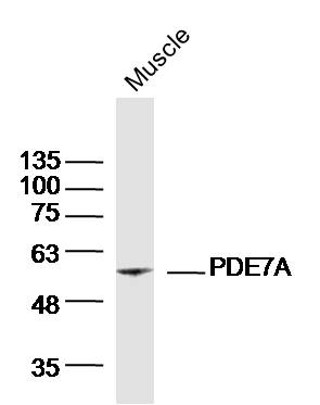
| Background Descriptions | Phosphodiesterases (PDE, also designated cyclic nucleotide phosphodiesterase) are important for the downregulation of the intracellular level of the second messenger cyclic adenosine monophosphate (cAMP) by hydrolyzing cAMP to 5'AMP. Phosphodiesterase type 3 isoforms, PDE3A and 3B, are expressed primarily in cardiovascular tissue and adipose tissue, respectively. PDE3A, is found in myocardium and platelets and PDE3B is found in lymphocytes. The PDE7A1 (HCP1) isozyme and the PDE7A2 proteins, alternate splice products of PDE7A, are highly expressed in skeletal muscle. PDE7B is most highly expressed in pancreas. The PDE family contains proteins that serve tissue-specific roles in regulation of lipolysis, glycogenolysis, myocardial contractility, and smooth muscle relaxation. |
| Gene ID | 5150 |
|---|---|
| Other Names | High affinity 3', 5'-cyclic-AMP phosphodiesterase 7A, 3.1.4.53, HCP1, TM22, cAMP-specific phosphodiesterase 7A, PDE7A {ECO:0000303|PubMed:9195912, ECO:0000312|HGNC:HGNC:8791} |
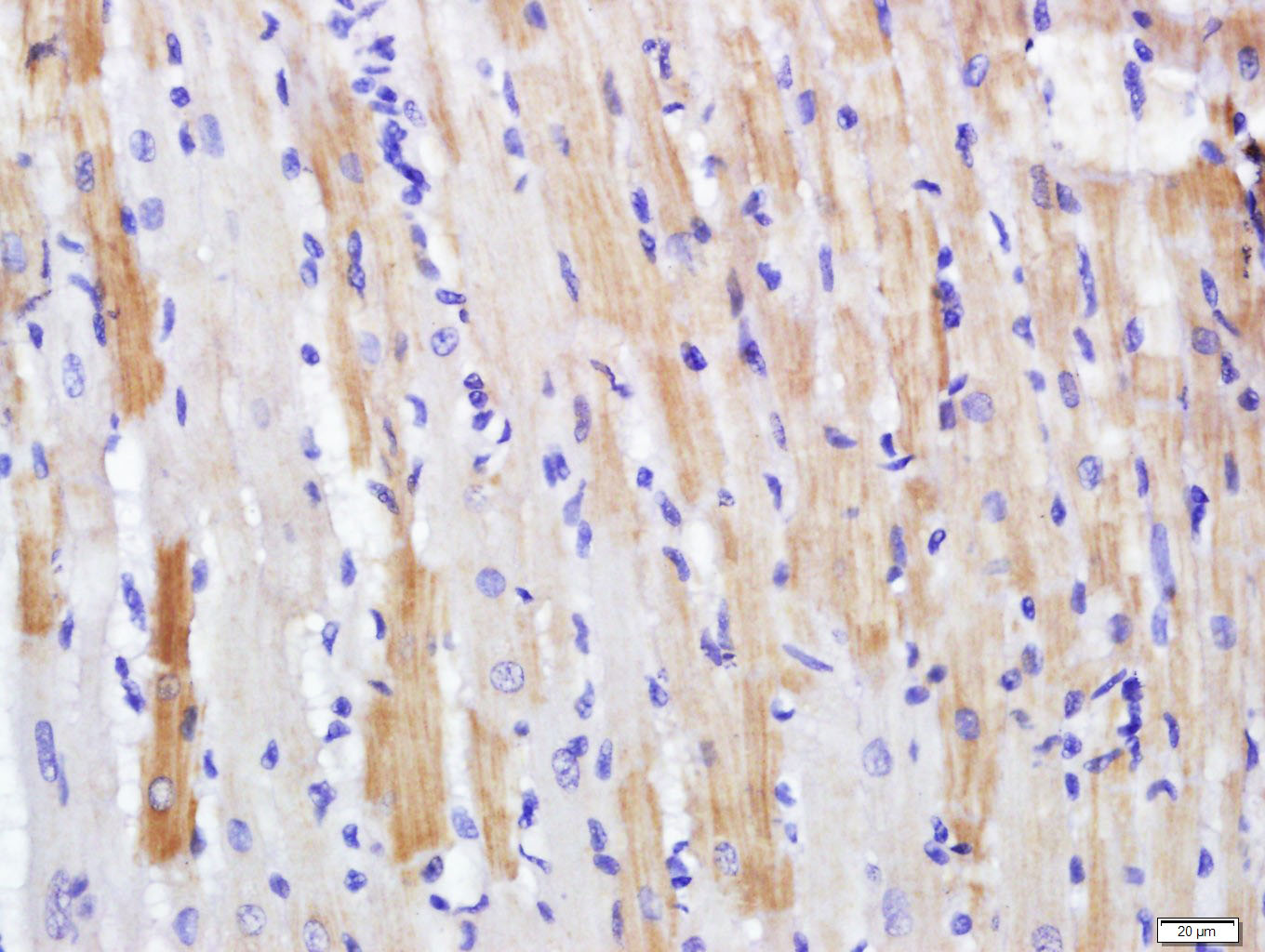
| Target/Specificity | PDE7A1 is found at high levels in skeletal muscle and at low levels in a variety of tissues including brain and heart. It is expressed as well in two T-cell lines. PDE7A2 is found abundantly in skeletal muscle and at low levels in heart. |
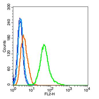
| Dilution | WB=1:500-2000,IHC-P=1:100-500,IHC-F=1:100-500,IF=1:100-500,Flow-Cyt=1 µg/Test |
| Storage | Store at -20 °C for one year. Avoid repeated freeze/thaw cycles. When reconstituted in sterile pH 7.4 0.01M PBS or diluent of antibody the antibody is stable for at least two weeks at 2-4 °C. |
| Name | PDE7A {ECO:0000303|PubMed:9195912, ECO:0000312|HGNC:HGNC:8791} |
|---|---|
| Function | Hydrolyzes the second messenger cAMP, which is a key regulator of many important physiological processes (PubMed:19350606, PubMed:8389765, PubMed:9195912). May have a role in muscle signal transduction (PubMed:9195912). |
| Cellular Location | [Isoform PDE7A1]: Cytoplasm, cytosol. Note=PDE7A1 (57 kDa) is located mostly to soluble cellular fractions. |
| Tissue Location | [Isoform PDE7A1]: Found at high levels in skeletal muscle and at low levels in a variety of tissues including brain and heart (PubMed:9195912). It is expressed as well in two T-cell lines (PubMed:9195912). |
For Research Use Only. Not For Use In Diagnostic Procedures.
Provided below are standard protocols that you may find useful for product applications.
BACKGROUND
Phosphodiesterases (PDE, also designated cyclic nucleotide phosphodiesterase) are important for the downregulation of the intracellular level of the second messenger cyclic adenosine monophosphate (cAMP) by hydrolyzing cAMP to 5'AMP. Phosphodiesterase type 3 isoforms, PDE3A and 3B, are expressed primarily in cardiovascular tissue and adipose tissue, respectively. PDE3A, is found in myocardium and platelets and PDE3B is found in lymphocytes. The PDE7A1 (HCP1) isozyme and the PDE7A2 proteins, alternate splice products of PDE7A, are highly expressed in skeletal muscle. PDE7B is most highly expressed in pancreas. The PDE family contains proteins that serve tissue-specific roles in regulation of lipolysis, glycogenolysis, myocardial contractility, and smooth muscle relaxation.
终于等到您。ABCEPTA(百远生物)抗体产品。
点击下方“我要评价 ”按钮提交您的反馈信息,您的反馈和评价是我们最宝贵的财富之一,
我们将在1-3个工作日内处理您的反馈信息。
如有疑问,联系:0512-88856768 tech-china@abcepta.com.


