


 癌症的基本特征包括细胞增殖、血管生成、迁移、凋亡逃避机制和细胞永生等。找到癌症发生过程中这些通路的关键标记物和对应的抗体用于检测至关重要。
 为您推荐一个泛素化位点预测神器——泛素化分析工具,可以为您的蛋白的泛素化位点作出预测和评分。
 细胞自噬受体图形绘图工具为你的蛋白的细胞受体结合位点作出预测和评分,识别结合到自噬通路中的蛋白是非常重要的,便于让我们理解自噬在正常生理、病理过程中的作用,如发育、细胞分化、神经退化性疾病、压力条件下、感染和癌症。
ATBF1 Rabbit pAb
ATBF1 Rabbit pAb
- 产品详情
- 实验流程
- 背景知识
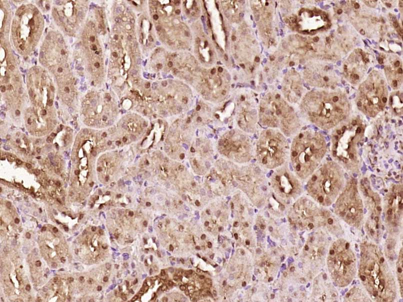
Application
| IHC-P, IHC-F, IF |
|---|---|
| Primary Accession | Q15911 |
| Reactivity | Rat |
| Predicted | Human, Mouse, Chicken, Dog, Horse, Rabbit, Sheep |
| Host | Rabbit |
| Clonality | Polyclonal |
| Calculated MW | 404419 Da |
| Physical State | Liquid |
| Immunogen | KLH conjugated synthetic peptide derived from human ATBF1 |
| Epitope Specificity | 301-400/3703 |
| Isotype | IgG |
| Purity | affinity purified by Protein A |
| Buffer | 0.01M TBS (pH7.4) with 1% BSA, 0.02% Proclin300 and 50% Glycerol. |
| SUBCELLULAR LOCATION | Nuclear |
| SIMILARITY | Contains 22 C2H2-type zinc fingers. Contains 4 homeobox DNA-binding domains. |
| SUBUNIT | Interacts with FNBP3 (By similarity). Interacts with PIAS3. |
| Post-translational modifications | Phosphorylated upon DNA damage, probably by ATM or ATR. |
| Important Note | This product as supplied is intended for research use only, not for use in human, therapeutic or diagnostic applications. |
| Background Descriptions | AT-motif binding factor 1 (ATBF1) binds to the AT-rich core sequence element in the human a-fetoprotein enhancer (1). Alternative splicing generates the ATBF1-A and ATBF1-B (2,3). While ATBF1-A contains a 920-amino acid extension at the N-terminus, both ATBF1-A and ATBF1-B contain 4 DNA-binding homeobox domains (2,3). Additionally, ATBF1-A contains 23 zinc finger motifs while ATBF1-B contains 18 zinc finger motifs (1–3). The N-terminal extension unique to ATBF1-A has transcriptional repressor activity (4). In the small intestine, ATBF1-A inhibits expression of the brushborder enzyme aminopeptidase-N through direct binding to the AT motif element (5). Besides functioning in transcription regulation, ATBF1 also functions in ATPase activity (6). ATPase activity associated with ATBF1-A is DNA/RNA-dependent and requires both homeobox domains and zinc finger motifs (6). ATBF1 is highly expressed in spleen and brain tissues (7). The gene encoding human ATBF1 maps to chromosome 16q22.3-q23.1 (8). |
| Gene ID | 463 |
|---|---|
| Other Names | Zinc finger homeobox protein 3, AT motif-binding factor 1, AT-binding transcription factor 1, Alpha-fetoprotein enhancer-binding protein, Zinc finger homeodomain protein 3, ZFH-3, ZFHX3, ATBF1, C16orf47 {ECO:0000312|HGNC:HGNC:777} |
| Target/Specificity | Not found in normal gastric mucosa but found in gastric carcinoma cells (at protein level). |
| Dilution | IHC-P=1:100-500,IHC-F=1:100-500,IF=1:100-500 |
| Storage | Store at -20 °C for one year. Avoid repeated freeze/thaw cycles. When reconstituted in sterile pH 7.4 0.01M PBS or diluent of antibody the antibody is stable for at least two weeks at 2-4 °C. |
| Name | ZFHX3 |
|---|---|
| Synonyms | ATBF1, C16orf47 {ECO:0000312|HGNC:HGNC:7 |
| Function | Transcriptional regulator which can act as an activator or a repressor. Inhibits the enhancer element of the AFP gene by binding to its AT-rich core sequence. In concert with SMAD-dependent TGF-beta signaling can repress the transcription of AFP via its interaction with SMAD2/3 (PubMed:25105025). Regulates the circadian locomotor rhythms via transcriptional activation of neuropeptidergic genes which are essential for intercellular synchrony and rhythm amplitude in the suprachiasmatic nucleus (SCN) of the brain (By similarity). Regulator of myoblasts differentiation through the binding to the AT-rich sequence of MYF6 promoter and promoter repression (PubMed:11312261). Down-regulates the MUC5AC promoter in gastric cancer (PubMed:17330845). In association with RUNX3, up-regulates CDKN1A promoter activity following TGF-beta stimulation (PubMed:20599712). Inhibits estrogen receptor (ESR1) function by selectively competing with coactivator NCOA3 for binding to ESR1 in ESR1-positive breast cancer cells (PubMed:20720010). |
| Cellular Location | Nucleus. Cytoplasm Note=Translocates from the cytoplasm to the nucleus following TGF-beta stimulation. Expressed in nuclear body (NB)-like dots in the nucleus some of which overlap or closely associate with PML body |
| Tissue Location | Not found in normal gastric mucosa but found in gastric carcinoma cells (at protein level). Expression is higher in ER- positive breast tumors than ER-negative breast tumors (at protein level). |
For Research Use Only. Not For Use In Diagnostic Procedures.
Provided below are standard protocols that you may find useful for product applications.
BACKGROUND
AT-motif binding factor 1 (ATBF1) binds to the AT-rich core sequence element in the human a-fetoprotein enhancer (1). Alternative splicing generates the ATBF1-A and ATBF1-B (2,3). While ATBF1-A contains a 920-amino acid extension at the N-terminus, both ATBF1-A and ATBF1-B contain 4 DNA-binding homeobox domains (2,3). Additionally, ATBF1-A contains 23 zinc finger motifs while ATBF1-B contains 18 zinc finger motifs (1–3). The N-terminal extension unique to ATBF1-A has transcriptional repressor activity (4). In the small intestine, ATBF1-A inhibits expression of the brushborder enzyme aminopeptidase-N through direct binding to the AT motif element (5). Besides functioning in transcription regulation, ATBF1 also functions in ATPase activity (6). ATPase activity associated with ATBF1-A is DNA/RNA-dependent and requires both homeobox domains and zinc finger motifs (6). ATBF1 is highly expressed in spleen and brain tissues (7). The gene encoding human ATBF1 maps to chromosome 16q22.3-q23.1 (8).
终于等到您。ABCEPTA(百远生物)抗体产品。
点击下方“我要评价 ”按钮提交您的反馈信息,您的反馈和评价是我们最宝贵的财富之一,
我们将在1-3个工作日内处理您的反馈信息。
如有疑问,联系:0512-88856768 tech-china@abcepta.com.


