


 癌症的基本特征包括细胞增殖、血管生成、迁移、凋亡逃避机制和细胞永生等。找到癌症发生过程中这些通路的关键标记物和对应的抗体用于检测至关重要。
 为您推荐一个泛素化位点预测神器——泛素化分析工具,可以为您的蛋白的泛素化位点作出预测和评分。
 细胞自噬受体图形绘图工具为你的蛋白的细胞受体结合位点作出预测和评分,识别结合到自噬通路中的蛋白是非常重要的,便于让我们理解自噬在正常生理、病理过程中的作用,如发育、细胞分化、神经退化性疾病、压力条件下、感染和癌症。
KIAA1598 Rabbit pAb
KIAA1598 Rabbit pAb
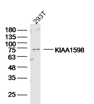
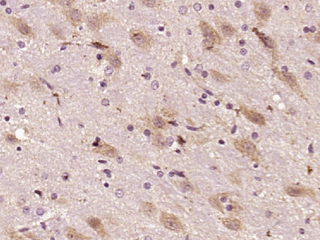
- 产品详情
- 实验流程
- 背景知识
Application 
| WB, IHC-P, IHC-F, IF |
|---|---|
| Primary Accession | A0MZ66 |
| Reactivity | Rat, Human |
| Predicted | Pig, Mouse, Horse |
| Host | Rabbit |
| Clonality | Polyclonal |
| Calculated MW | 71640 Da |
| Physical State | Liquid |
| Immunogen | KLH conjugated synthetic peptide derived from human KIAA1598 |
| Epitope Specificity | 21-130/631 |
| Isotype | IgG |
| Purity | affinity purified by Protein A |
| Buffer | 0.01M TBS (pH7.4) with 1% BSA, 0.02% Proclin300 and 50% Glycerol. |
| SUBCELLULAR LOCATION | Cell projection. |
| Important Note | This product as supplied is intended for research use only, not for use in human, therapeutic or diagnostic applications. |
| Background Descriptions | Shootin1 is a 631 amino acid protein that belongs to the shootin family. The shootin1 protein contains three coiled-coil domains, a proline-rich region and interacts with RUFY3. Shootin1 is involved in the generation of internal asymmetric signals required for neuronal polarization. The shootin1 protein acts upstream of PI3K (phosphoinositide 3-kinase), by being required for spatially localized PI3K activity. By accumulating asymmetrically in a single neurite before polarization, shootin1 leads to axon induction for polarization, additionally the absence of shootin1 from the nascent axon's siblings by competition prevents the formation of surplus axons. Existing as seven alternatively spliced isoforms, the shootin1 gene is conserved in chimpanzee, dog, mouse, rat, chicken and zebrafish, and maps to human chromosome 10q25.3. |
| Gene ID | 57698 |
|---|---|
| Other Names | Shootin-1 {ECO:0000312|HGNC:HGNC:29319}, Shootin1, SHTN1 (HGNC:29319), KIAA1598 |
| Dilution | WB=1:500-2000,IHC-P=1:100-500,IHC-F=1:100-500,IF=1:100-500 |
| Storage | Store at -20 °C for one year. Avoid repeated freeze/thaw cycles. When reconstituted in sterile pH 7.4 0.01M PBS or diluent of antibody the antibody is stable for at least two weeks at 2-4 °C. |
For Research Use Only. Not For Use In Diagnostic Procedures.
| Name | SHTN1 (HGNC:29319) |
|---|---|
| Synonyms | KIAA1598 |
| Function | Involved in the generation of internal asymmetric signals required for neuronal polarization and neurite outgrowth. Mediates netrin-1-induced F-actin-substrate coupling or 'clutch engagement' within the axon growth cone through activation of CDC42, RAC1 and PAK1- dependent signaling pathway, thereby converting the F-actin retrograde flow into traction forces, concomitantly with filopodium extension and axon outgrowth. Plays a role in cytoskeletal organization by regulating the subcellular localization of phosphoinositide 3-kinase (PI3K) activity at the axonal growth cone. Also plays a role in regenerative neurite outgrowth. In the developing cortex, cooperates with KIF20B to promote both the transition from the multipolar to the bipolar stage and the radial migration of cortical neurons from the ventricular zone toward the superficial layer of the neocortex. Involved in the accumulation of phosphatidylinositol 3,4,5-trisphosphate (PIP3) in the growth cone of primary hippocampal neurons. |
| Cellular Location | Perikaryon {ECO:0000250|UniProtKB:Q8K2Q9}. Cell projection, axon {ECO:0000250|UniProtKB:Q8K2Q9}. Cell projection, growth cone {ECO:0000250|UniProtKB:Q8K2Q9}. Cytoplasm, cytoskeleton {ECO:0000250|UniProtKB:Q8K2Q9}. Cell projection, filopodium {ECO:0000250|UniProtKB:A0MZ67}. Cell projection, lamellipodium {ECO:0000250|UniProtKB:A0MZ67}. Note=Localizes in multiple growth cones at neurite tips before the neuronal symmetry-breaking step. Accumulates in growth cones of a single nascent axon in a neurite length-dependent manner during the neuronal symmetry-breaking step; when absent from the nascent axon's siblings, probably due to competitive transport, prevents the formation of surplus axons. Transported anterogradely from the soma to the axon growth cone in an actin and myosin-dependent manner and passively diffuses back to the cell bodies. Colocalized with L1CAM in close apposition with actin filaments in filopodia and lamellipodia of axonal growth cones in hippocampal neurons. Exhibits retrograde movements in filopodia and lamellopodia of axonal growth cones. Colocalized with KIF20B along microtubules to the tip of the growing cone in primary hippocampal neurons. Recruited to the growth cone of developing axon in a KIF20B- and microtubule-dependent manner {ECO:0000250|UniProtKB:A0MZ67, ECO:0000250|UniProtKB:Q8K2Q9} |
Research Areas
Application Protocols
Provided below are standard protocols that you may find useful for product applications.
BACKGROUND
This product as supplied is intended for research use only, not for use in human, therapeutic or diagnostic applications.

终于等到您。ABCEPTA(百远生物)抗体产品。
点击下方“我要评价 ”按钮提交您的反馈信息,您的反馈和评价是我们最宝贵的财富之一,
我们将在1-3个工作日内处理您的反馈信息。
如有疑问,联系:0512-88856768 tech-china@abcepta.com.


