


 癌症的基本特征包括细胞增殖、血管生成、迁移、凋亡逃避机制和细胞永生等。找到癌症发生过程中这些通路的关键标记物和对应的抗体用于检测至关重要。
 为您推荐一个泛素化位点预测神器——泛素化分析工具,可以为您的蛋白的泛素化位点作出预测和评分。
 细胞自噬受体图形绘图工具为你的蛋白的细胞受体结合位点作出预测和评分,识别结合到自噬通路中的蛋白是非常重要的,便于让我们理解自噬在正常生理、病理过程中的作用,如发育、细胞分化、神经退化性疾病、压力条件下、感染和癌症。
PLEKHG5 Rabbit pAb
PLEKHG5 Rabbit pAb
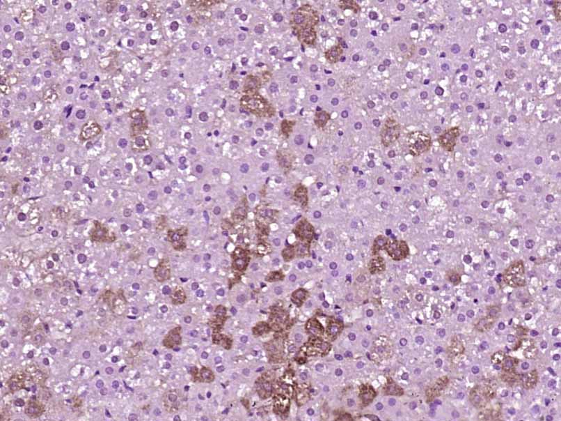
- 产品详情
- 实验流程
- 背景知识
Application 
| IHC-P, IHC-F, IF |
|---|---|
| Primary Accession | O94827 |
| Reactivity | Rat, Mouse |
| Predicted | Pig, Dog, Human, Rabbit, Horse |
| Host | Rabbit |
| Clonality | Polyclonal |
| Calculated MW | 111231 Da |
| Physical State | Liquid |
| Immunogen | KLH conjugated synthetic peptide derived from human PLEKHG5 |
| Epitope Specificity | 951-1062/1062 |
| Isotype | IgG |
| Purity | affinity purified by Protein A |
| Buffer | 0.01M TBS (pH7.4) with 1% BSA, 0.02% Proclin300 and 50% Glycerol. |
| SUBCELLULAR LOCATION | Cytoplasm. Cytoplasm, perinuclear region. Cell junction. Cell projection, lamellipodium. Note=Predominantly cytoplasmic, however when cells are stimulated found in perinuclear regions. Localized at cell-cell junctions in quiescent endothelial cells, it relocalizes to cytoplasmic vesicle and the leading edge of lamellipodia in migrating endothelial cells. |
| DISEASE | Distal spinal muscular atrophy, autosomal recessive, 4 (DSMA4) [MIM:611067]: A neuromuscular disorder. Distal spinal muscular atrophy, also known as distal hereditary motor neuronopathy, represents a heterogeneous group of neuromuscular disorders caused by selective degeneration of motor neurons in the anterior horn of the spinal cord, without sensory deficit in the posterior horn. The overall clinical picture consists of a classical distal muscular atrophy syndrome in the legs without clinical sensory loss. The disease starts with weakness and wasting of distal muscles of the anterior tibial and peroneal compartments of the legs. Later on, weakness and atrophy may expand to the proximal muscles of the lower limbs and/or to the distal upper limbs. DSMA4 is characterized by childhood onset, generalized muscle weakness and atrophy with denervation and normal sensation. Bulbar symptoms and pyramidal signs are absent. Note=The disease is caused by mutations affecting the gene represented in this entry. Charcot-Marie-Tooth disease, recessive, intermediate type, C (CMTRIC) [MIM:615376]: A form of Charcot-Marie-Tooth disease, a disorder of the peripheral nervous system, characterized by progressive weakness and atrophy, initially of the peroneal muscles and later of the distal muscles of the arms. Recessive intermediate forms of Charcot-Marie-Tooth disease are characterized by clinical and pathologic features intermediate between demyelinating and axonal peripheral neuropathies, and motor median nerve conduction velocities ranging from 25 to 45 m/sec. Note=The disease is caused by mutations affecting the gene represented in this entry. |
| Important Note | This product as supplied is intended for research use only, not for use in human, therapeutic or diagnostic applications. |
| Background Descriptions | This gene encodes a protein that activates the nuclear factor kappa B (NFKB1) signaling pathway. Mutations in this gene are associated with autosomal recessive distal spinal muscular atrophy. Multiple transcript variants encoding different isoforms have been found for this gene. [provided by RefSeq, May 2012] |
| Gene ID | 57449 |
|---|---|
| Other Names | Pleckstrin homology domain-containing family G member 5, PH domain-containing family G member 5, Guanine nucleotide exchange factor 720, GEF720, PLEKHG5, KIAA0720 |
| Dilution | IHC-P=1:100-500,IHC-F=1:100-500,IF=1:100-500 |
| Storage | Store at -20 °C for one year. Avoid repeated freeze/thaw cycles. When reconstituted in sterile pH 7.4 0.01M PBS or diluent of antibody the antibody is stable for at least two weeks at 2-4 °C. |
For Research Use Only. Not For Use In Diagnostic Procedures.
| Name | PLEKHG5 |
|---|---|
| Synonyms | KIAA0720 |
| Function | Functions as a guanine exchange factor (GEF) for RAB26 and thus regulates autophagy of synaptic vesicles in axon terminal of motoneurons (By similarity). Involved in the control of neuronal cell differentiation (PubMed:11704860). Plays a role in angiogenesis through regulation of endothelial cells chemotaxis. Also affects the migration, adhesion, and matrix/bone degradation in macrophages and osteoclasts (PubMed:23777631). |
| Cellular Location | Cytoplasm {ECO:0000250|UniProtKB:Q66T02}. Cytoplasm, perinuclear region {ECO:0000250|UniProtKB:Q66T02}. Cell membrane {ECO:0000250|UniProtKB:Q66T02}. Cell junction {ECO:0000250|UniProtKB:Q66T02}. Cell projection, lamellipodium {ECO:0000250|UniProtKB:Q66T02}. Note=Predominantly cytoplasmic, however when endothelial cells are stimulated with lysophosphatidic acid, PLEKHG5 is found in perinuclear regions and at the cell membrane Localizes at cell-cell junctions in quiescent endothelial cells, and relocalizes to cytoplasmic vesicle and the leading edge of lamellipodia in migrating endothelial cells. {ECO:0000250|UniProtKB:Q66T02} |
| Tissue Location | Predominantly expressed in the peripheral nervous system and brain. Highest expression is observed in heart, lung, kidney, testis and moderate expression is present in spleen, pancreas, skeletal muscle, ovary and liver. Weakly expressed in glioblastoma (GBM) cell lines. |
Research Areas
Application Protocols
Provided below are standard protocols that you may find useful for product applications.
BACKGROUND
This product as supplied is intended for research use only, not for use in human, therapeutic or diagnostic applications.

终于等到您。ABCEPTA(百远生物)抗体产品。
点击下方“我要评价 ”按钮提交您的反馈信息,您的反馈和评价是我们最宝贵的财富之一,
我们将在1-3个工作日内处理您的反馈信息。
如有疑问,联系:0512-88856768 tech-china@abcepta.com.


