


 癌症的基本特征包括细胞增殖、血管生成、迁移、凋亡逃避机制和细胞永生等。找到癌症发生过程中这些通路的关键标记物和对应的抗体用于检测至关重要。
 为您推荐一个泛素化位点预测神器——泛素化分析工具,可以为您的蛋白的泛素化位点作出预测和评分。
 细胞自噬受体图形绘图工具为你的蛋白的细胞受体结合位点作出预测和评分,识别结合到自噬通路中的蛋白是非常重要的,便于让我们理解自噬在正常生理、病理过程中的作用,如发育、细胞分化、神经退化性疾病、压力条件下、感染和癌症。
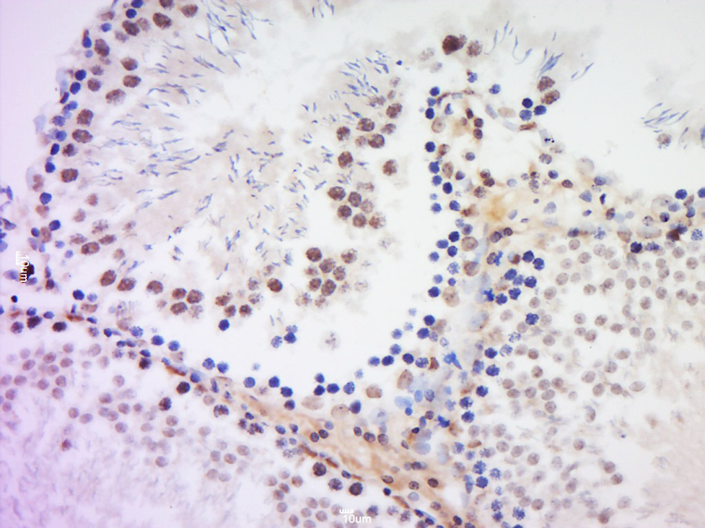
HSH2D Rabbit pAb
HSH2D Rabbit pAb
- 产品详情
- 实验流程
- 背景知识
Application
| IHC-P, IHC-F, IF |
|---|---|
| Primary Accession | Q96JZ2 |
| Reactivity | Rat |
| Predicted | Human, Mouse, Dog, Horse, Sheep |
| Host | Rabbit |
| Clonality | Polyclonal |
| Calculated MW | 39002 Da |
| Physical State | Liquid |
| Immunogen | KLH conjugated synthetic peptide derived from human HSH2D |
| Epitope Specificity | 21-120/352 |
| Isotype | IgG |
| Purity | affinity purified by Protein A |
| Buffer | 0.01M TBS (pH7.4) with 1% BSA, 0.02% Proclin300 and 50% Glycerol. |
| SUBCELLULAR LOCATION | Cytoplasm. Nucleus. |
| SIMILARITY | Contains 1 SH2 domain. |
| Post-translational modifications | May be phosphorylated by FES and ACK1. |
| Important Note | This product as supplied is intended for research use only, not for use in human, therapeutic or diagnostic applications. |
| Background Descriptions | HSH2 is a 352 amino acid nuclear and cytoplasmic protein that is predominantly expressed in spleen and hematopoietic cells, such as peripheral blood leukocytes, and weakly expressed in prostate, thymus, heart, small intestine and placenta. Containing an SH2 domain, four PXXP polyproline sequences and two possible tyrosine phosphorylation sites, HSH2 interacts with tyrosine kinases Fes and ACK. Considered an adaptor protein, HSH2 participates in tyrosine kinase signaling and may be involved in the regulation of cytokine signaling and cytoskeletal reorganization in hematopoietic cells. HSH2 may also act to attenuate apoptosis through modulating the apoptotic response by promoting mitochondrial stability. HSH2 exists as two alternatively spliced isoforms and is encoded by a gene located on human chromosome 19p13.11. |
| Gene ID | 84941 |
|---|---|
| Other Names | Hematopoietic SH2 domain-containing protein, Hematopoietic SH2 protein, Adaptor in lymphocytes of unknown function X, HSH2D, ALX |
| Target/Specificity | Predominantly expressed in spleen and hematopoietic cells such as peripheral blood leukocytes and weakly expressed in prostate, thymus, heart, small intestine and placenta. |
| Dilution | IHC-P=1:100-500,IHC-F=1:100-500,IF=1:100-500 |
| Storage | Store at -20 °C for one year. Avoid repeated freeze/thaw cycles. When reconstituted in sterile pH 7.4 0.01M PBS or diluent of antibody the antibody is stable for at least two weeks at 2-4 °C. |
| Name | HSH2D |
|---|---|
| Synonyms | ALX |
| Function | May be a modulator of the apoptotic response through its ability to affect mitochondrial stability (By similarity). Adapter protein involved in tyrosine kinase and CD28 signaling. Seems to affect CD28-mediated activation of the RE/AP element of the interleukin-2 promoter. |
| Cellular Location | Cytoplasm. Nucleus. |
| Tissue Location | Predominantly expressed in spleen and hematopoietic cells such as peripheral blood leukocytes and weakly expressed in prostate, thymus, heart, small intestine and placenta |
For Research Use Only. Not For Use In Diagnostic Procedures.
Provided below are standard protocols that you may find useful for product applications.
BACKGROUND
HSH2 is a 352 amino acid nuclear and cytoplasmic protein that is predominantly expressed in spleen and hematopoietic cells, such as peripheral blood leukocytes, and weakly expressed in prostate, thymus, heart, small intestine and placenta. Containing an SH2 domain, four PXXP polyproline sequences and two possible tyrosine phosphorylation sites, HSH2 interacts with tyrosine kinases Fes and ACK. Considered an adaptor protein, HSH2 participates in tyrosine kinase signaling and may be involved in the regulation of cytokine signaling and cytoskeletal reorganization in hematopoietic cells. HSH2 may also act to attenuate apoptosis through modulating the apoptotic response by promoting mitochondrial stability. HSH2 exists as two alternatively spliced isoforms and is encoded by a gene located on human chromosome 19p13.11.
终于等到您。ABCEPTA(百远生物)抗体产品。
点击下方“我要评价 ”按钮提交您的反馈信息,您的反馈和评价是我们最宝贵的财富之一,
我们将在1-3个工作日内处理您的反馈信息。
如有疑问,联系:0512-88856768 tech-china@abcepta.com.


