


 癌症的基本特征包括细胞增殖、血管生成、迁移、凋亡逃避机制和细胞永生等。找到癌症发生过程中这些通路的关键标记物和对应的抗体用于检测至关重要。
 为您推荐一个泛素化位点预测神器——泛素化分析工具,可以为您的蛋白的泛素化位点作出预测和评分。
 细胞自噬受体图形绘图工具为你的蛋白的细胞受体结合位点作出预测和评分,识别结合到自噬通路中的蛋白是非常重要的,便于让我们理解自噬在正常生理、病理过程中的作用,如发育、细胞分化、神经退化性疾病、压力条件下、感染和癌症。
PSMD9 Rabbit pAb
PSMD9 Rabbit pAb
- 产品详情
- 实验流程
- 背景知识
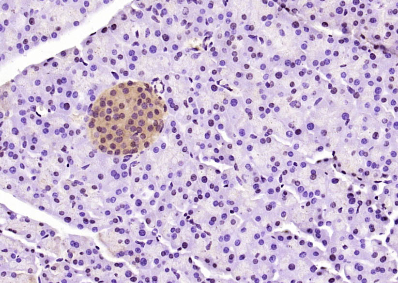
Application
| IHC-P, IHC-F, IF |
|---|---|
| Primary Accession | O00233 |
| Reactivity | Mouse, Rat |
| Predicted | Human, Chicken, Dog, Pig, Horse, Rabbit, Guinea Pig |
| Host | Rabbit |
| Clonality | Polyclonal |
| Calculated MW | 24682 Da |
| Physical State | Liquid |
| Immunogen | KLH conjugated synthetic peptide derived from human PSMD9 |
| Epitope Specificity | 65-150/223 |
| Isotype | IgG |
| Purity | affinity purified by Protein A |
| Buffer | 0.01M TBS (pH7.4) with 1% BSA, 0.02% Proclin300 and 50% Glycerol. |
| SIMILARITY | Belongs to the proteasome subunit p27 family.Contains 1 PDZ (DHR) domain. |
| SUBUNIT | Interacts with PSMC3. Part of a transient complex (modulator) containing PSMD9, PSMC6 and PSMC3 formed during the assembly of the 26S proteasome. |
| Important Note | This product as supplied is intended for research use only, not for use in human, therapeutic or diagnostic applications. |
| Background Descriptions | The 26S proteasome is a multicatalytic proteinase complex with a highly ordered structure composed of 2 complexes, a 20S core and a 19S regulator. The 20S core is composed of 4 rings of 28 non-identical subunits; 2 rings are composed of 7 alpha subunits and 2 rings are composed of 7 beta subunits. The 19S regulator is composed of a base, which contains 6 ATPase subunits and 2 non-ATPase subunits, and a lid, which contains up to 10 non-ATPase subunits. Proteasomes are distributed throughout eukaryotic cells at a high concentration and cleave peptides in an ATP/ubiquitin-dependent process in a non-lysosomal pathway. An essential function of a modified proteasome, the immunoproteasome, is the processing of class I MHC peptides. This gene encodes a non-ATPase subunit of the 19S regulator. Three transcript variants encoding two different isoforms have been found for this gene. [provided by RefSeq, May 2012] |
| Gene ID | 5715 |
|---|---|
| Other Names | 26S proteasome non-ATPase regulatory subunit 9, 26S proteasome regulatory subunit p27, PSMD9 |
| Target/Specificity | Expressed in all tissues tested, highly expressed in liver and kidney. |
| Dilution | IHC-P=1:100-500,IHC-F=1:100-500,IF=1:100-500 |
| Storage | Store at -20 °C for one year. Avoid repeated freeze/thaw cycles. When reconstituted in sterile pH 7.4 0.01M PBS or diluent of antibody the antibody is stable for at least two weeks at 2-4 °C. |
| Name | PSMD9 |
|---|---|
| Function | Acts as a chaperone during the assembly of the 26S proteasome, specifically of the base subcomplex of the PA700/19S regulatory complex (RC). During the base subcomplex assembly is part of an intermediate PSMD9:PSMC6:PSMC3 module, also known as modulator trimer complex; PSMD9 is released during the further base assembly process. |
| Tissue Location | Expressed in all tissues tested, highly expressed in liver and kidney |
For Research Use Only. Not For Use In Diagnostic Procedures.
Provided below are standard protocols that you may find useful for product applications.
BACKGROUND
The 26S proteasome is a multicatalytic proteinase complex with a highly ordered structure composed of 2 complexes, a 20S core and a 19S regulator. The 20S core is composed of 4 rings of 28 non-identical subunits; 2 rings are composed of 7 alpha subunits and 2 rings are composed of 7 beta subunits. The 19S regulator is composed of a base, which contains 6 ATPase subunits and 2 non-ATPase subunits, and a lid, which contains up to 10 non-ATPase subunits. Proteasomes are distributed throughout eukaryotic cells at a high concentration and cleave peptides in an ATP/ubiquitin-dependent process in a non-lysosomal pathway. An essential function of a modified proteasome, the immunoproteasome, is the processing of class I MHC peptides. This gene encodes a non-ATPase subunit of the 19S regulator. Three transcript variants encoding two different isoforms have been found for this gene. [provided by RefSeq, May 2012]
终于等到您。ABCEPTA(百远生物)抗体产品。
点击下方“我要评价 ”按钮提交您的反馈信息,您的反馈和评价是我们最宝贵的财富之一,
我们将在1-3个工作日内处理您的反馈信息。
如有疑问,联系:0512-88856768 tech-china@abcepta.com.


