


 癌症的基本特征包括细胞增殖、血管生成、迁移、凋亡逃避机制和细胞永生等。找到癌症发生过程中这些通路的关键标记物和对应的抗体用于检测至关重要。
 为您推荐一个泛素化位点预测神器——泛素化分析工具,可以为您的蛋白的泛素化位点作出预测和评分。
 细胞自噬受体图形绘图工具为你的蛋白的细胞受体结合位点作出预测和评分,识别结合到自噬通路中的蛋白是非常重要的,便于让我们理解自噬在正常生理、病理过程中的作用,如发育、细胞分化、神经退化性疾病、压力条件下、感染和癌症。
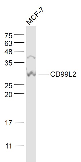
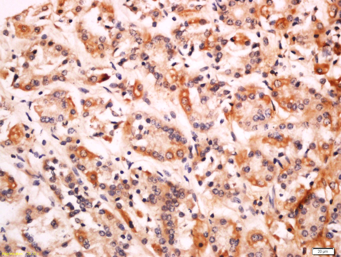
CD99L2 Rabbit pAb
CD99L2 Rabbit pAb
- 产品详情
- 实验流程
- 背景知识
Application
| WB, IHC-P, IHC-F, IF |
|---|---|
| Primary Accession | Q8TCZ2 |
| Reactivity | Human |
| Host | Rabbit |
| Clonality | Polyclonal |
| Calculated MW | 27986 Da |
| Physical State | Liquid |
| Immunogen | KLH conjugated synthetic peptide derived from human CD99L2 |
| Epitope Specificity | 81-187/262 |
| Isotype | IgG |
| Purity | affinity purified by Protein A |
| Buffer | 0.01M TBS (pH7.4) with 1% BSA, 0.02% Proclin300 and 50% Glycerol. |
| SUBCELLULAR LOCATION | Cell membrane. Cell junction. |
| SIMILARITY | Belongs to the CD99 family. |
| Post-translational modifications | O-glycosylated. |
| Important Note | This product as supplied is intended for research use only, not for use in human, therapeutic or diagnostic applications. |
| Background Descriptions | May function as a homophilic adhesion molecule. Functions in leukocyte-endothelial cell interactions during leukocyte extravasation, and in particular, at the diapedesis step. Does not seem to be involved in docking of leukocytes to the vessel wall or in lymphocyte diapedesis. |
| Gene ID | 83692 |
|---|---|
| Other Names | CD99 antigen-like protein 2, MIC2-like protein 1, CD99, CD99L2, MIC2L1 |
| Target/Specificity | Expressed in brain, heart, lung, liver, spleen, kidney, stomach, small intestine, skeletal muscle, ovary, thymus, testis and uterus. Lower expression seen in thymus. Expressed in E18 uterus and placenta. |
| Dilution | WB=1:500-2000,IHC-P=1:100-500,IHC-F=1:100-500,IF=1:100-500 |
| Storage | Store at -20 °C for one year. Avoid repeated freeze/thaw cycles. When reconstituted in sterile pH 7.4 0.01M PBS or diluent of antibody the antibody is stable for at least two weeks at 2-4 °C. |
| Name | CD99L2 |
|---|---|
| Synonyms | MIC2L1 |
| Function | Plays a role in a late step of leukocyte extravasation helping cells to overcome the endothelial basement membrane. Acts at the same site as, but independently of, PECAM1 (By similarity). Homophilic adhesion molecule, but these interactions may not be required for cell aggregation (By similarity). |
| Cellular Location | Cell membrane; Single-pass type I membrane protein; Extracellular side. Cell junction. Secreted |
| Tissue Location | Detected in cerebrospinal fluid (at protein level) (PubMed:25326458). Expressed in many tissues, with low expression in thymus. |
Research Areas
For Research Use Only. Not For Use In Diagnostic Procedures.
Application Protocols
Provided below are standard protocols that you may find useful for product applications.
BACKGROUND
May function as a homophilic adhesion molecule. Functions in leukocyte-endothelial cell interactions during leukocyte extravasation, and in particular, at the diapedesis step. Does not seem to be involved in docking of leukocytes to the vessel wall or in lymphocyte diapedesis.
终于等到您。ABCEPTA(百远生物)抗体产品。
点击下方“我要评价 ”按钮提交您的反馈信息,您的反馈和评价是我们最宝贵的财富之一,
我们将在1-3个工作日内处理您的反馈信息。
如有疑问,联系:0512-88856768 tech-china@abcepta.com.


