


 癌症的基本特征包括细胞增殖、血管生成、迁移、凋亡逃避机制和细胞永生等。找到癌症发生过程中这些通路的关键标记物和对应的抗体用于检测至关重要。
 为您推荐一个泛素化位点预测神器——泛素化分析工具,可以为您的蛋白的泛素化位点作出预测和评分。
 细胞自噬受体图形绘图工具为你的蛋白的细胞受体结合位点作出预测和评分,识别结合到自噬通路中的蛋白是非常重要的,便于让我们理解自噬在正常生理、病理过程中的作用,如发育、细胞分化、神经退化性疾病、压力条件下、感染和癌症。
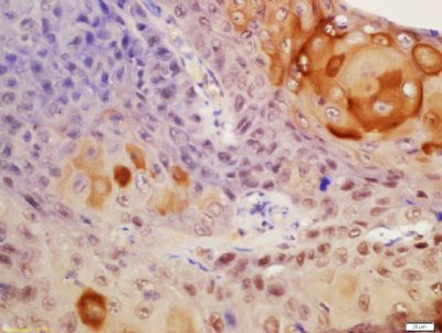
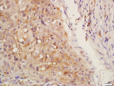
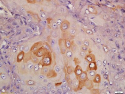
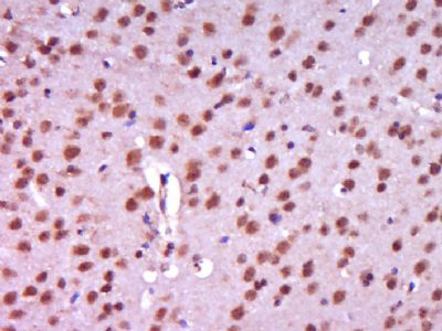
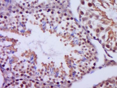
Centrin 2 Polyclonal Antibody
Purified Rabbit Polyclonal Antibody (Pab)
- 产品详情
- 实验流程
Application
| IHC-P, IHC-F, IF, ICC, E |
|---|---|
| Primary Accession | P41208 |
| Reactivity | Rat, Pig, Bovine |
| Host | Rabbit |
| Clonality | Polyclonal |
| Calculated MW | 19738 Da |
| Physical State | Liquid |
| Immunogen | KLH conjugated synthetic peptide derived from human Centrin 2 |
| Epitope Specificity | 1-100/172 |
| Isotype | IgG |
| Purity | affinity purified by Protein A |
| Buffer | 0.01M TBS (pH7.4) with 1% BSA, 0.02% Proclin300 and 50% Glycerol. |
| SUBCELLULAR LOCATION | Cytoplasm > cytoskeleton > centrosome > centriole. Nucleus. Centrosome of S-phase, interphase and mitotic cells. |
| SIMILARITY | Belongs to the centrin family. Contains 4 EF-hand domains. |
| Important Note | This product as supplied is intended for research use only, not for use in human, therapeutic or diagnostic applications. |
| Background Descriptions | Caltractin belongs to a family of calcium-binding proteins and is a structural component of the centrosome. The high level of conservation from algae to humans and its association with the centrosome suggested that caltractin plays a fundamental role in the structure and function of the microtubule-organizing center, possibly required for the proper duplication and segregation of the centrosome. [provided by RefSeq, Jul 2008] |
| Gene ID | 1069 |
|---|---|
| Other Names | Centrin-2, Caltractin isoform 1, CETN2, CALT, CEN2 |
| Dilution | IHC-P=1:100-500,IHC-F=1:100-500,ICC=1:100-500,IF=1:100-500,ELISA=1:5000-10000 |
| Format | 0.01M TBS(pH7.4) with 1% BSA, 0.09% (W/V) sodium azide and 50% Glyce |
| Storage | Store at -20 °C for one year. Avoid repeated freeze/thaw cycles. When reconstituted in sterile pH 7.4 0.01M PBS or diluent of antibody the antibody is stable for at least two weeks at 2-4 °C. |
| Name | CETN2 |
|---|---|
| Synonyms | CALT, CEN2 |
| Function | Plays a fundamental role in microtubule organizing center structure and function. Required for centriole duplication and correct spindle formation. Has a role in regulating cytokinesis and genome stability via cooperation with CALM1 and CCP110. The XPC complex is proposed to represent the first factor bound at the sites of DNA damage and together with other core recognition factors, XPA, RPA and the TFIIH complex, is part of the pre-incision (or initial recognition) complex. The XPC complex recognizes a wide spectrum of damaged DNA characterized by distortions of the DNA helix such as single-stranded loops, mismatched bubbles or single-stranded overhangs. The orientation of XPC complex binding appears to be crucial for inducing a productive NER. XPC complex is proposed to recognize and to interact with unpaired bases on the undamaged DNA strand which is followed by recruitment of the TFIIH complex and subsequent scanning for lesions in the opposite strand in a 5'-to-3' direction by the NER machinery. Cyclobutane pyrimidine dimers (CPDs) which are formed upon UV-induced DNA damage esacpe detection by the XPC complex due to a low degree of structural perurbation. Instead they are detected by the UV-DDB complex which in turn recruits and cooperates with the XPC complex in the respective DNA repair. |
| Cellular Location | Cytoplasm, cytoskeleton, microtubule organizing center, centrosome. Cytoplasm, cytoskeleton, microtubule organizing center, centrosome, centriole. Nucleus envelope. Nucleus, nuclear pore complex. Nucleus. Note=Localizes to the inner scaffold in the central region of centrioles and to the distal end of centrioles. |
Research Areas
For Research Use Only. Not For Use In Diagnostic Procedures.
Application Protocols
Provided below are standard protocols that you may find useful for product applications.
终于等到您。ABCEPTA(百远生物)抗体产品。
点击下方“我要评价 ”按钮提交您的反馈信息,您的反馈和评价是我们最宝贵的财富之一,
我们将在1-3个工作日内处理您的反馈信息。
如有疑问,联系:0512-88856768 tech-china@abcepta.com.
¥ 1,500.00
Cat# AP55318


