


 癌症的基本特征包括细胞增殖、血管生成、迁移、凋亡逃避机制和细胞永生等。找到癌症发生过程中这些通路的关键标记物和对应的抗体用于检测至关重要。
 为您推荐一个泛素化位点预测神器——泛素化分析工具,可以为您的蛋白的泛素化位点作出预测和评分。
 细胞自噬受体图形绘图工具为你的蛋白的细胞受体结合位点作出预测和评分,识别结合到自噬通路中的蛋白是非常重要的,便于让我们理解自噬在正常生理、病理过程中的作用,如发育、细胞分化、神经退化性疾病、压力条件下、感染和癌症。
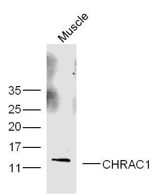
CHRAC1 Rabbit pAb
CHRAC1 Rabbit pAb
- 产品详情
- 实验流程
- 背景知识
Application
| WB |
|---|---|
| Primary Accession | Q9NRG0 |
| Reactivity | Human, Mouse |
| Predicted | Rat, Chicken, Sheep |
| Host | Rabbit |
| Clonality | Polyclonal |
| Calculated MW | 14711 Da |
| Physical State | Liquid |
| Immunogen | KLH conjugated synthetic peptide derived from human CHRAC1 |
| Epitope Specificity | 1-100/131 |
| Isotype | IgG |
| Purity | affinity purified by Protein A |
| Buffer | 0.01M TBS (pH7.4) with 1% BSA, 0.02% Proclin300 and 50% Glycerol. |
| SUBCELLULAR LOCATION | Nucleus. |
| Important Note | This product as supplied is intended for research use only, not for use in human, therapeutic or diagnostic applications. |
| Background Descriptions | DNA replication is initiated by the binding of initiation factors to the origin of replication. Nucleosomes inhibit access to the replication machinery at these origin sequences. Nucleosome remodeling factors increase the accessibility of nucleosomal DNA to transcriptional regulators (1). CHRAC15 and CHRAC17 are subunits of the nucleosomal remodeling factor CHRAC (chromatin accessibility complex),which increases the accessibility of nucleosomal DNA in an ATP-dependent manner (2). Unlike other known chromatin remodelling factors, CHRAC also functions during chromatin assembly by using ATP to convert irregular chromatin into a regular array of nucleosomes with even spacing (3). This conversion process occurs when CHRAC organizes randomly deposited histones into a regularly spaced array (4). In the presence of CHRAC, the nucleosomal ATPase ISWI catalyses several ATP-dependent transitions of chromatin structure (5). |
| Gene ID | 54108 |
|---|---|
| Other Names | Chromatin accessibility complex protein 1, CHRAC-1, Chromatin accessibility complex 15 kDa protein, CHRAC-15, HuCHRAC15, DNA polymerase epsilon subunit p15, CHRAC1, CHRAC15 |
| Target/Specificity | Expressed in all tissues tested, including, heart, brain, placenta, lung, liver, skeletal muscle, kidney and pancreas. |
| Dilution | WB=1:500-2000 |
| Storage | Store at -20 °C for one year. Avoid repeated freeze/thaw cycles. When reconstituted in sterile pH 7.4 0.01M PBS or diluent of antibody the antibody is stable for at least two weeks at 2-4 °C. |
| Name | CHRAC1 |
|---|---|
| Synonyms | CHRAC15 |
| Function | Forms a complex with DNA polymerase epsilon subunit POLE3 and binds naked DNA, which is then incorporated into chromatin, aided by the nucleosome remodeling activity of ISWI/SNF2H and ACF1. Does not enhance nucleosome sliding activity of the ACF-5 ISWI chromatin remodeling complex (PubMed:14759371). |
| Cellular Location | Nucleus. |
| Tissue Location | Expressed in heart, brain, placenta, lung, liver, skeletal muscle, kidney and pancreas. |
For Research Use Only. Not For Use In Diagnostic Procedures.
Provided below are standard protocols that you may find useful for product applications.
BACKGROUND
DNA replication is initiated by the binding of initiation factors to the origin of replication. Nucleosomes inhibit access to the replication machinery at these origin sequences. Nucleosome remodeling factors increase the accessibility of nucleosomal DNA to transcriptional regulators (1). CHRAC15 and CHRAC17 are subunits of the nucleosomal remodeling factor CHRAC (chromatin accessibility complex),which increases the accessibility of nucleosomal DNA in an ATP-dependent manner (2). Unlike other known chromatin remodelling factors, CHRAC also functions during chromatin assembly by using ATP to convert irregular chromatin into a regular array of nucleosomes with even spacing (3). This conversion process occurs when CHRAC organizes randomly deposited histones into a regularly spaced array (4). In the presence of CHRAC, the nucleosomal ATPase ISWI catalyses several ATP-dependent transitions of chromatin structure (5).
终于等到您。ABCEPTA(百远生物)抗体产品。
点击下方“我要评价 ”按钮提交您的反馈信息,您的反馈和评价是我们最宝贵的财富之一,
我们将在1-3个工作日内处理您的反馈信息。
如有疑问,联系:0512-88856768 tech-china@abcepta.com.


