


 癌症的基本特征包括细胞增殖、血管生成、迁移、凋亡逃避机制和细胞永生等。找到癌症发生过程中这些通路的关键标记物和对应的抗体用于检测至关重要。
 为您推荐一个泛素化位点预测神器——泛素化分析工具,可以为您的蛋白的泛素化位点作出预测和评分。
 细胞自噬受体图形绘图工具为你的蛋白的细胞受体结合位点作出预测和评分,识别结合到自噬通路中的蛋白是非常重要的,便于让我们理解自噬在正常生理、病理过程中的作用,如发育、细胞分化、神经退化性疾病、压力条件下、感染和癌症。
CHST7 Rabbit pAb
CHST7 Rabbit pAb
- 产品详情
- 实验流程
- 背景知识
Application
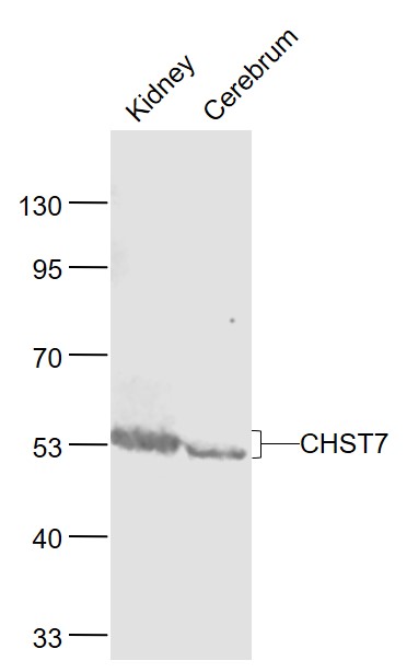
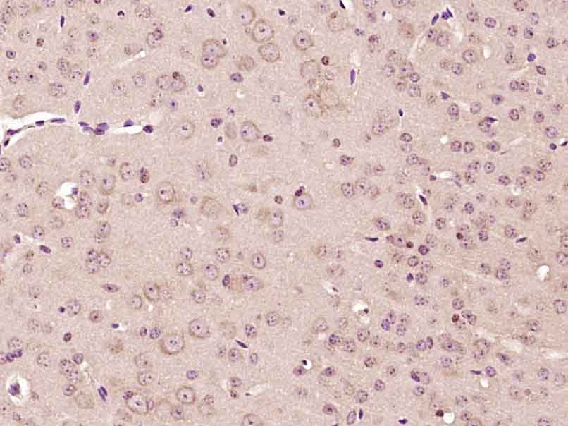
| WB, IHC-P, IHC-F, IF |
|---|---|
| Primary Accession | Q9NS84 |
| Reactivity | Mouse |
| Predicted | Human, Rat, Dog, Pig, Rabbit, Sheep |
| Host | Rabbit |
| Clonality | Polyclonal |
| Calculated MW | 54266 Da |
| Physical State | Liquid |
| Immunogen | KLH conjugated synthetic peptide derived from human CHST7 |
| Epitope Specificity | 101-200/486 |
| Isotype | IgG |
| Purity | affinity purified by Protein A |
| Buffer | 0.01M TBS (pH7.4) with 1% BSA, 0.02% Proclin300 and 50% Glycerol. |
| SUBCELLULAR LOCATION | Golgi apparatus membrane; Single-pass type II membrane protein |
| SIMILARITY | Belongs to the sulfotransferase 1 family. Gal/GlcNAc/GalNAc subfamily. |
| Important Note | This product as supplied is intended for research use only, not for use in human, therapeutic or diagnostic applications. |
| Background Descriptions | This gene encodes a DNA topoisomerase, an enzyme that controls and alters the topologic states of DNA during transcription. This enzyme catalyzes the transient breaking and rejoining of a single strand of DNA which allows the strands to pass through one another, thus reducing the number of supercoils and altering the topology of DNA. This enzyme forms a complex with BLM which functions in the regulation of recombination in somatic cells. [provided by RefSeq, Jul 2008] |
| Gene ID | 56548 |
|---|---|
| Other Names | Carbohydrate sulfotransferase 7, 2.8.2.-, 2.8.2.17, Chondroitin 6-sulfotransferase 2, C6ST-2, Galactose/N-acetylglucosamine/N-acetylglucosamine 6-O-sulfotransferase 5, GST-5, N-acetylglucosamine 6-O-sulfotransferase 4, GlcNAc6ST-4, Gn6st-4, CHST7 |
| Target/Specificity | Widely expressed. Highly expressed in heart, spleen, liver and ovary. Expressed at lower level in brain, placenta, thyroid, spinal cord and peripheral blood leukocytes. Not expressed in adult skin. |
| Dilution | WB=1:500-2000,IHC-P=1:100-500,IHC-F=1:100-500,IF=1:100-500 |
| Storage | Store at -20 °C for one year. Avoid repeated freeze/thaw cycles. When reconstituted in sterile pH 7.4 0.01M PBS or diluent of antibody the antibody is stable for at least two weeks at 2-4 °C. |
| Name | CHST7 |
|---|---|
| Function | Sulfotransferase that utilizes 3'-phospho-5'-adenylyl sulfate (PAPS) as sulfonate donor to catalyze the transfer of sulfate to position 6 of non-reducing N-acetylglucosamine (GlcNAc) residues. Preferentially acts on mannose-linked GlcNAc. Also able to catalyze the transfer of sulfate to position 6 of the N-acetylgalactosamine (GalNAc) residue of chondroitin. Also acts on core 2 mucin-type oligosaccharide and N-acetyllactosamine oligomer with a lower efficiency. Has weak or no activity toward keratan sulfate and oligosaccharides containing the Galbeta1-4GlcNAc. Catalyzes 6-O-sulfation of beta-benzyl GlcNAc but not alpha- or beta-benzyl GalNAc. |
| Cellular Location | Golgi apparatus membrane; Single- pass type II membrane protein |
| Tissue Location | Widely expressed. Highly expressed in heart, spleen, liver and ovary. Expressed at lower level in brain, placenta, thyroid, spinal cord and peripheral blood leukocytes. Not expressed in adult skin. |
For Research Use Only. Not For Use In Diagnostic Procedures.
Provided below are standard protocols that you may find useful for product applications.
BACKGROUND
This gene encodes a DNA topoisomerase, an enzyme that controls and alters the topologic states of DNA during transcription. This enzyme catalyzes the transient breaking and rejoining of a single strand of DNA which allows the strands to pass through one another, thus reducing the number of supercoils and altering the topology of DNA. This enzyme forms a complex with BLM which functions in the regulation of recombination in somatic cells. [provided by RefSeq, Jul 2008]
终于等到您。ABCEPTA(百远生物)抗体产品。
点击下方“我要评价 ”按钮提交您的反馈信息,您的反馈和评价是我们最宝贵的财富之一,
我们将在1-3个工作日内处理您的反馈信息。
如有疑问,联系:0512-88856768 tech-china@abcepta.com.


