


 癌症的基本特征包括细胞增殖、血管生成、迁移、凋亡逃避机制和细胞永生等。找到癌症发生过程中这些通路的关键标记物和对应的抗体用于检测至关重要。
 为您推荐一个泛素化位点预测神器——泛素化分析工具,可以为您的蛋白的泛素化位点作出预测和评分。
 细胞自噬受体图形绘图工具为你的蛋白的细胞受体结合位点作出预测和评分,识别结合到自噬通路中的蛋白是非常重要的,便于让我们理解自噬在正常生理、病理过程中的作用,如发育、细胞分化、神经退化性疾病、压力条件下、感染和癌症。
CRB1 Rabbit pAb
CRB1 Rabbit pAb
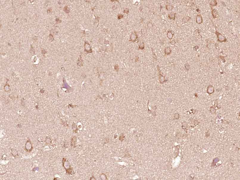
- 产品详情
- 实验流程
- 背景知识
Application 
| IHC-P, IHC-F, IF |
|---|---|
| Primary Accession | P82279 |
| Reactivity | Human |
| Host | Rabbit |
| Clonality | Polyclonal |
| Calculated MW | 154183 Da |
| Physical State | Liquid |
| Immunogen | KLH conjugated synthetic peptide derived from human CRB1 |
| Epitope Specificity | 301-400/1406 |
| Isotype | IgG |
| Purity | affinity purified by Protein A |
| Buffer | 0.01M TBS (pH7.4) with 1% BSA, 0.02% Proclin300 and 50% Glycerol. |
| SUBCELLULAR LOCATION | Secreted and Apical cell membrane. Distributed at the apical membrane of all retinal epithelial cells. Located in the apical membrane of the adherens junction in outer limiting membrane (OLM) of the retina. |
| DISEASE | Note=CRB1 mutations have been found in various retinal dystrophies, chronic and disabling disorders of visual function. They predominantly involve the posterior portion of the ocular fundus, due to degeneration in the sensory layer of the retina, retinal pigment epithelium, Bruch membrane, choroid, or a combination of these tissues. Onset of inherited retinal dystrophies is painless, bilateral and typically progressive. Most people experience gradual peripheral vision loss or tunnel vision, and difficulties with poor illumination and night vision. Central vision is usually unaffected, so the person may still be able to read. However, it can also deteriorate to cause total blindness. Examples of retinal dystrophies are retinitis pigmentosa, Leber congenital amaurosis, cone-rod dystrophy among others. Defects in CRB1 are the cause of retinitis pigmentosa type 12 (RP12) [MIM:600105]. A retinal dystrophy belonging to the group of pigmentary retinopathies. Retinitis pigmentosa is characterized by retinal pigment deposits visible on fundus examination and primary loss of rod photoreceptor cells, followed by secondary loss of cone photoreceptors. Patients typically have night vision blindness and loss of midperipheral visual field. As their condition progresses, they lose their far peripheral visual field and eventually central vision as well. RP12 is an autosomal recessive severe form oFTen manifesting in early childhood. Patients experiment progressive visual field loss with severe visual impairment before the age of twenty. Some patients have a preserved paraarteriolar retinal pigment epithelium (PPRPE) and hypermetropia. Defects in CRB1 are the cause of Leber congenital amaurosis type 8 (LCA8) [MIM:613835]. LCA designates a clinically and genetically heterogeneous group of childhood retinal degenerations, generally inherited in an autosomal recessive manner. Affected infants have little or no retinal photoreceptor function as tested by electroretinography. LCA represents the most common genetic cause of congenital visual impairment in infants and children. Defects in CRB1 are the cause of pigmented paravenous chorioretinal atrophy (PPCRA) [MIM:172870]. PPCRA is an unusual retinal degeneration characterized by accumulation of pigmentation along retinal veins. PPCRA is dominantly inherited, but exhibited variable expressivity. Males are more likely to exhibit a severe phenotype, whereas females may remain virtually asymptomatic even in later years. The PPCRA phenotype is associated with a mutation in CRB1 gene which is likely to affect the structure of the CRB1 protein. |
| Important Note | This product as supplied is intended for research use only, not for use in human, therapeutic or diagnostic applications. |
| Background Descriptions | This gene encodes a protein which is similar to the Drosophila crumbs protein and localizes to the inner segment of mammalian photoreceptors. In Drosophila crumbs localizes to the stalk of the fly photoreceptor and may be a component of the molecular scaffold that controls proper development of polarity in the eye. Mutations in this gene are associated with a severe form of retinitis pigmentosa, RP12, and with Leber congenital amaurosis. Alternate splicing results in multiple transcript variants, some protein coding and some non-protein coding.[provided by RefSeq, Apr 2012] |
| Gene ID | 23418 |
|---|---|
| Other Names | Protein crumbs homolog 1, CRB1 (HGNC:2343) |
| Dilution | IHC-P=1:100-500,IHC-F=1:100-500,IF=1:100-500 |
| Storage | Store at -20 °C for one year. Avoid repeated freeze/thaw cycles. When reconstituted in sterile pH 7.4 0.01M PBS or diluent of antibody the antibody is stable for at least two weeks at 2-4 °C. |
For Research Use Only. Not For Use In Diagnostic Procedures.
| Name | CRB1 (HGNC:2343) |
|---|---|
| Function | Plays a role in photoreceptor morphogenesis in the retina (By similarity). May maintain cell polarization and adhesion (By similarity). |
| Cellular Location | [Isoform 1]: Apical cell membrane {ECO:0000250|UniProtKB:Q8VHS2}; Single-pass type I membrane protein. Secreted. Cell projection, cilium, photoreceptor outer segment {ECO:0000250|UniProtKB:Q8VHS2} Photoreceptor inner segment {ECO:0000250|UniProtKB:Q8VHS2} |
| Tissue Location | Preferential expression in retina, also expressed in brain, testis, fetal brain and fetal eye (PubMed:15914641) Expressed at the outer limiting membrane and apical to adherens junctions in the retina (PubMed:15914641) |
Research Areas
Application Protocols
Provided below are standard protocols that you may find useful for product applications.
BACKGROUND
This product as supplied is intended for research use only, not for use in human, therapeutic or diagnostic applications.

终于等到您。ABCEPTA(百远生物)抗体产品。
点击下方“我要评价 ”按钮提交您的反馈信息,您的反馈和评价是我们最宝贵的财富之一,
我们将在1-3个工作日内处理您的反馈信息。
如有疑问,联系:0512-88856768 tech-china@abcepta.com.


