


 癌症的基本特征包括细胞增殖、血管生成、迁移、凋亡逃避机制和细胞永生等。找到癌症发生过程中这些通路的关键标记物和对应的抗体用于检测至关重要。
 为您推荐一个泛素化位点预测神器——泛素化分析工具,可以为您的蛋白的泛素化位点作出预测和评分。
 细胞自噬受体图形绘图工具为你的蛋白的细胞受体结合位点作出预测和评分,识别结合到自噬通路中的蛋白是非常重要的,便于让我们理解自噬在正常生理、病理过程中的作用,如发育、细胞分化、神经退化性疾病、压力条件下、感染和癌症。
PIEZO1 Rabbit pAb
PIEZO1 Rabbit pAb
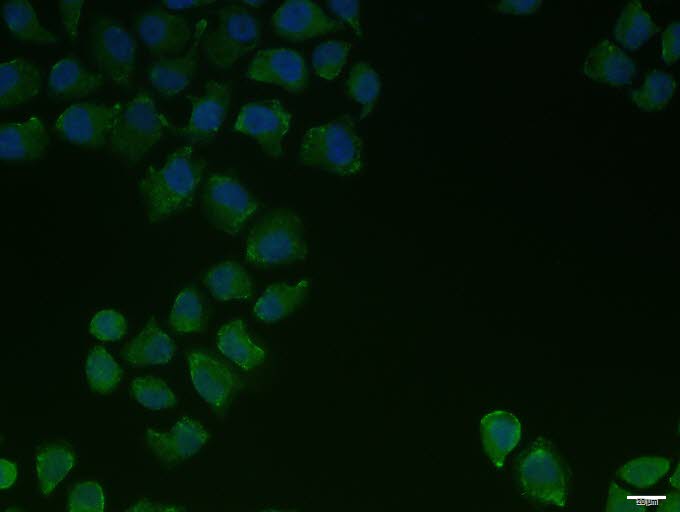
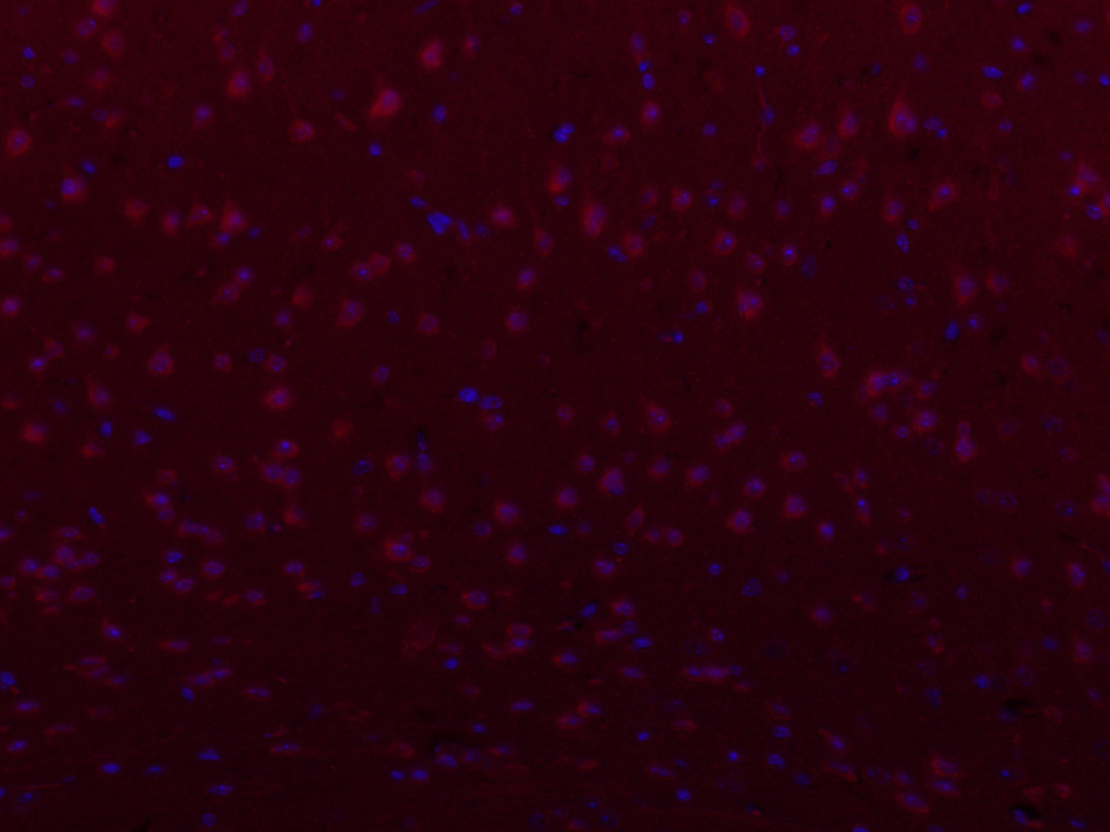
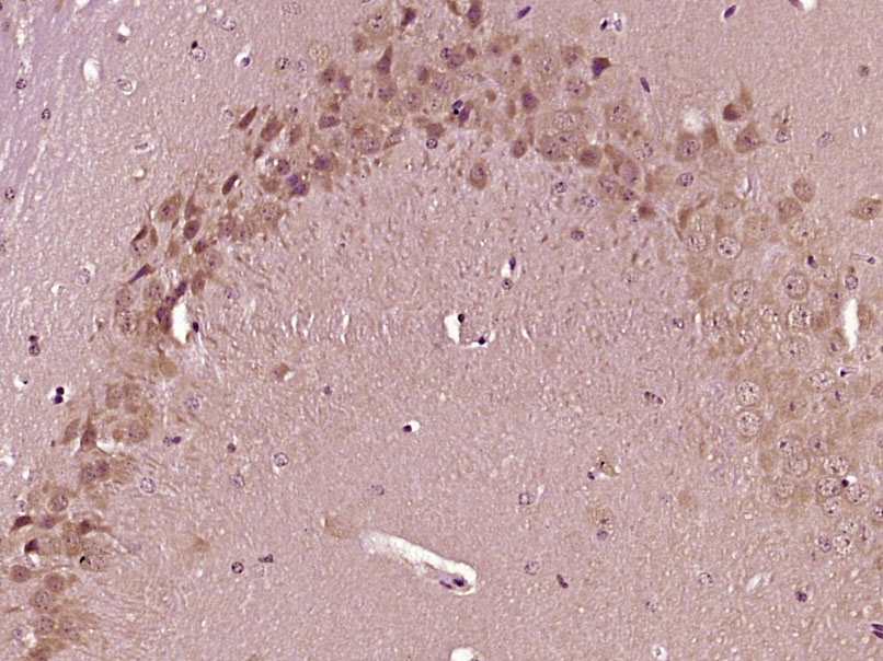
- 产品详情
- 实验流程
- 背景知识
Application 
| IHC-P, IHC-F, IF |
|---|---|
| Primary Accession | Q92508 |
| Reactivity | Human, Mouse |
| Predicted | Rat, Dog, Sheep |
| Host | Rabbit |
| Clonality | Polyclonal |
| Calculated MW | 286790 Da |
| Physical State | Liquid |
| Immunogen | KLH conjugated synthetic peptide derived from human FAM38A |
| Epitope Specificity | 2451-2521/2521 |
| Isotype | IgG |
| Purity | affinity purified by Protein A |
| Buffer | 0.01M TBS (pH7.4) with 1% BSA, 0.02% Proclin300 and 50% Glycerol. |
| SUBCELLULAR LOCATION | Endoplasmic reticulum membrane. Endoplasmic reticulum-Golgi intermediate compartment membrane. Cell membrane. |
| Important Note | This product as supplied is intended for research use only, not for use in human, therapeutic or diagnostic applications. |
| Background Descriptions | The protein encoded by this gene is a mechanically-activated ion channel that links mechanical forces to biological signals. The encoded protein contains 36 transmembrane domains and functions as a homotetramer. Defects in this gene have been associated with dehydrated hereditary stomatocytosis. [provided by RefSeq, Jul 2015] |
| Gene ID | 9780 |
|---|---|
| Other Names | Piezo-type mechanosensitive ion channel component 1, Membrane protein induced by beta-amyloid treatment, Mib, Protein FAM38A, PIEZO1 (HGNC:28993), FAM38A, KIAA0233 |
| Dilution | IHC-P=1:100-500,IHC-F=1:100-500,ICC/IF=1:100-500,IF=1:100-500 |
| Storage | Store at -20 °C for one year. Avoid repeated freeze/thaw cycles. When reconstituted in sterile pH 7.4 0.01M PBS or diluent of antibody the antibody is stable for at least two weeks at 2-4 °C. |
For Research Use Only. Not For Use In Diagnostic Procedures.
| Name | PIEZO1 (HGNC:28993) |
|---|---|
| Synonyms | FAM38A, KIAA0233 |
| Function | Pore-forming subunit of the mechanosensitive non-specific cation Piezo channel required for rapidly adapting mechanically activated (MA) currents and has a key role in sensing touch and tactile pain (PubMed:23479567, PubMed:23695678, PubMed:25955826, PubMed:37590348). Piezo channels are homotrimeric three-blade propeller-shaped structures that utilize a cap-motion and plug-and- latch mechanism to gate their ion-conducting pathways (PubMed:37590348). Generates currents characterized by a linear current-voltage relationship that are sensitive to ruthenium red and gadolinium (By similarity). Conductance to monovalent alkali ions is highest for K(+), intermediate for Na(+) and lowest for Li(+) (PubMed:25955826). Divalent ions except for Mn(2+) permeate the channel but more slowly than the monovalent ions and they also reduce K(+) currents (PubMed:25955826). Plays a key role in epithelial cell adhesion by maintaining integrin activation through R-Ras recruitment to the ER, most probably in its activated state, and subsequent stimulation of calpain signaling (PubMed:20016066). In inner ear hair cells, PIEZO1/2 subunits may constitute part of the mechanotransducer (MET) non-selective cation channel complex where they may act as pore- forming ion-conducting component in the complex (By similarity). In the kidney, may contribute to the detection of intraluminal pressure changes and to urine flow sensing (By similarity). Acts as a shear- stress sensor that promotes endothelial cell organization and alignment in the direction of blood flow through calpain activation (PubMed:25119035). Plays a key role in blood vessel formation and vascular structure in both development and adult physiology (By similarity). Acts as a sensor of phosphatidylserine (PS) flipping at the plasma membrane and governs morphogenesis of muscle cells (By similarity). In myoblasts, flippase-mediated PS enrichment at the inner leaflet of plasma membrane triggers channel activation and Ca2+ influx followed by Rho GTPases signal transduction, leading to assembly of cortical actomyosin fibers and myotube formation (PubMed:29799007). |
| Cellular Location | Endoplasmic reticulum membrane; Multi-pass membrane protein {ECO:0000250|UniProtKB:E2JF22}. Endoplasmic reticulum-Golgi intermediate compartment membrane {ECO:0000250|UniProtKB:Q0KL00}. Cell membrane; Multi-pass membrane protein {ECO:0000250|UniProtKB:E2JF22}. Cell projection, lamellipodium membrane Note=In erythrocytes, located in the plasma membrane (PubMed:22529292, PubMed:23479567). Accumulates at the leading apical lamellipodia of endothelial cells in response to shear stress (PubMed:25119035) Colocalizes with F-actin and MYH9 at the actomyosin cortex in myoblasts. {ECO:0000250|UniProtKB:E2JF22, ECO:0000269|PubMed:22529292, ECO:0000269|PubMed:23479567, ECO:0000269|PubMed:25119035} |
| Tissue Location | Expressed in numerous tissues. In normal brain, expressed exclusively in neurons, not in astrocytes. In Alzheimer disease brains, expressed in about half of the activated astrocytes located around classical senile plaques. In Parkinson disease substantia nigra, not detected in melanin-containing neurons nor in activated astrocytes. Expressed in erythrocytes (at protein level) Expressed in myoblasts (at protein level) |
Research Areas
Application Protocols
Provided below are standard protocols that you may find useful for product applications.
BACKGROUND
This product as supplied is intended for research use only, not for use in human, therapeutic or diagnostic applications.

终于等到您。ABCEPTA(百远生物)抗体产品。
点击下方“我要评价 ”按钮提交您的反馈信息,您的反馈和评价是我们最宝贵的财富之一,
我们将在1-3个工作日内处理您的反馈信息。
如有疑问,联系:0512-88856768 tech-china@abcepta.com.


