


 癌症的基本特征包括细胞增殖、血管生成、迁移、凋亡逃避机制和细胞永生等。找到癌症发生过程中这些通路的关键标记物和对应的抗体用于检测至关重要。
 为您推荐一个泛素化位点预测神器——泛素化分析工具,可以为您的蛋白的泛素化位点作出预测和评分。
 细胞自噬受体图形绘图工具为你的蛋白的细胞受体结合位点作出预测和评分,识别结合到自噬通路中的蛋白是非常重要的,便于让我们理解自噬在正常生理、病理过程中的作用,如发育、细胞分化、神经退化性疾病、压力条件下、感染和癌症。
KIF20B Rabbit pAb
KIF20B Rabbit pAb
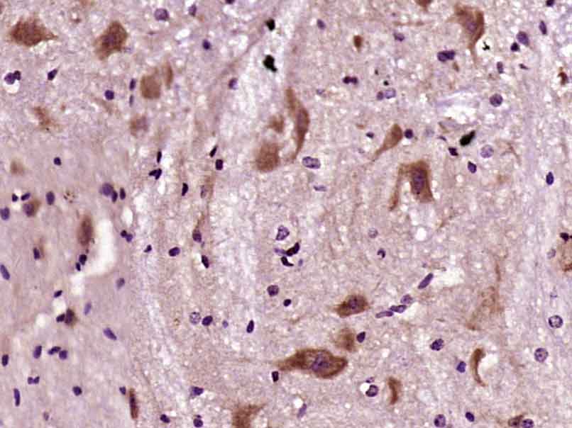
- 产品详情
- 实验流程
- 背景知识
Application 
| IHC-P, IHC-F, IF |
|---|---|
| Primary Accession | Q96Q89 |
| Reactivity | Mouse |
| Predicted | Rat, Pig, Dog, Human, Horse, Sheep |
| Host | Rabbit |
| Clonality | Polyclonal |
| Calculated MW | 210629 Da |
| Physical State | Liquid |
| Immunogen | KLH conjugated synthetic peptide derived from human KIF20B |
| Epitope Specificity | 1651-1750/1820 |
| Isotype | IgG |
| Purity | affinity purified by Protein A |
| Buffer | 0.01M TBS (pH7.4) with 1% BSA, 0.02% Proclin300 and 50% Glycerol. |
| SUBCELLULAR LOCATION | Cytoplasm. Nucleus. Cytoplasm > cytoskeleton > spindle. Localizes mainly in the nucleus during interphase although it is also detected in the cytoplasm without clear association with microtubules. A 2-3 fold expression increase is seen as cells progress from G1 to G2/M phase. During prophase and metaphase it is found throughout the cytoplasm and at anaphase accumulates at the midplan of the cell and forms a distinct band extending across the spindle midzone. At anaphase it is concentrated in the mid-body. |
| Important Note | This product as supplied is intended for research use only, not for use in human, therapeutic or diagnostic applications. |
| Background Descriptions | The Kinesins constitute a large family of microtubule-dependent motor proteins which are responsible for the distribution of numerous organelles, vesicles and macromolecular complexes throughout the cell. Individual Kinesin members play crucial roles in cell division, intracellular transport and membrane trafficking events, including endocytosis and transcytosis. MPP1 (M-phase phosphoprotein 1), also known as KIF20B (kinesin family member 20B), MPHOSPH1 or KRMP1, is a 1,820 amino acid protein that localizes to both the nucleus and the cytoplasm and contains one kinesin-motor domain. Expressed in kidney, brain, testis and ovary, MPP1 functions as a plus-end directed motor enzyme that interacts with Pin1 and is required for the completion of cytokinesis. MPP1, which exists as multiple alternatively spliced isoforms termed 1-5, is subject to post-translational phosphorylation, probably by ATM or ATR. |
| Gene ID | 9585 |
|---|---|
| Other Names | Kinesin-like protein KIF20B, Cancer/testis antigen 90, CT90, Kinesin family member 20B {ECO:0000312|HGNC:HGNC:7212}, Kinesin-related motor interacting with PIN1, M-phase phosphoprotein 1, MPP1, KIF20B (HGNC:7212) |
| Dilution | IHC-P=1:100-500,IHC-F=1:100-500,IF=1:100-500 |
| Storage | Store at -20 °C for one year. Avoid repeated freeze/thaw cycles. When reconstituted in sterile pH 7.4 0.01M PBS or diluent of antibody the antibody is stable for at least two weeks at 2-4 °C. |
For Research Use Only. Not For Use In Diagnostic Procedures.
| Name | KIF20B (HGNC:7212) |
|---|---|
| Function | Plus-end-directed motor enzyme that is required for completion of cytokinesis (PubMed:11470801, PubMed:12740395). Required for proper midbody organization and abscission in polarized cortical stem cells. Plays a role in the regulation of neuronal polarization by mediating the transport of specific cargos. Participates in the mobilization of SHTN1 and in the accumulation of PIP3 in the growth cone of primary hippocampal neurons in a tubulin and actin-dependent manner. In the developing telencephalon, cooperates with SHTN1 to promote both the transition from the multipolar to the bipolar stage and the radial migration of cortical neurons from the ventricular zone toward the superficial layer of the neocortex. Involved in cerebral cortex growth (By similarity). Acts as an oncogene for promoting bladder cancer cells proliferation, apoptosis inhibition and carcinogenic progression (PubMed:17409436). |
| Cellular Location | Nucleus. Cytoplasm, cytoskeleton, microtubule organizing center, centrosome. Nucleus, nucleolus. Nucleus, nucleoplasm. Cytoplasm, cytoskeleton, spindle. Cytoplasm, cytoskeleton, spindle pole. Midbody. Cell projection, axon {ECO:0000250|UniProtKB:Q80WE4}. Cell projection, growth cone {ECO:0000250|UniProtKB:Q80WE4}. Note=Localizes mainly in the nucleus during interphase although it is also detected in the cytoplasm without clear association with microtubules (PubMed:12740395). Localized to the central spindle during cytokinetic furrowing and with the midbody during abscission (PubMed:12740395, PubMed:17409436). A 2-3 fold expression increase is seen as cells progress from G1 to G2/M phase (PubMed:12740395). During prophase and metaphase it is found throughout the cytoplasm and at anaphase accumulates at the midplan of the cell and forms a distinct band extending across the spindle midzone (PubMed:12740395). At anaphase it is concentrated in the midbody (PubMed:12740395). Colocalized partially along microtubules in primary neurons. Colocalized with SHTN1 along microtubules to the tip of the growing cone in primary hippocampal neurons. Localized in midbodies between dividing radial progenitors in the ventricular zone (By similarity). Colocalized with PRC1 in the nucleus of bladder carcinoma cells at the interphase. Colocalized with PRC1 in bladder carcinoma cells at prophase, metaphase, early anaphase, at the midzone in late anaphase and at the contractile ring in telophase (PubMed:17409436). {ECO:0000250|UniProtKB:Q80WE4, ECO:0000269|PubMed:12740395, ECO:0000269|PubMed:17409436} |
| Tissue Location | Brain, ovary, kidney and testis (at protein level) (PubMed:12740395). Overexpressed in bladder cancer cells (at protein level) (PubMed:17409436). Expressed in testis. Overexpressed in bladder cancer cells (PubMed:17409436). |
Research Areas
Application Protocols
Provided below are standard protocols that you may find useful for product applications.
BACKGROUND
This product as supplied is intended for research use only, not for use in human, therapeutic or diagnostic applications.

终于等到您。ABCEPTA(百远生物)抗体产品。
点击下方“我要评价 ”按钮提交您的反馈信息,您的反馈和评价是我们最宝贵的财富之一,
我们将在1-3个工作日内处理您的反馈信息。
如有疑问,联系:0512-88856768 tech-china@abcepta.com.


