


 癌症的基本特征包括细胞增殖、血管生成、迁移、凋亡逃避机制和细胞永生等。找到癌症发生过程中这些通路的关键标记物和对应的抗体用于检测至关重要。
 为您推荐一个泛素化位点预测神器——泛素化分析工具,可以为您的蛋白的泛素化位点作出预测和评分。
 细胞自噬受体图形绘图工具为你的蛋白的细胞受体结合位点作出预测和评分,识别结合到自噬通路中的蛋白是非常重要的,便于让我们理解自噬在正常生理、病理过程中的作用,如发育、细胞分化、神经退化性疾病、压力条件下、感染和癌症。
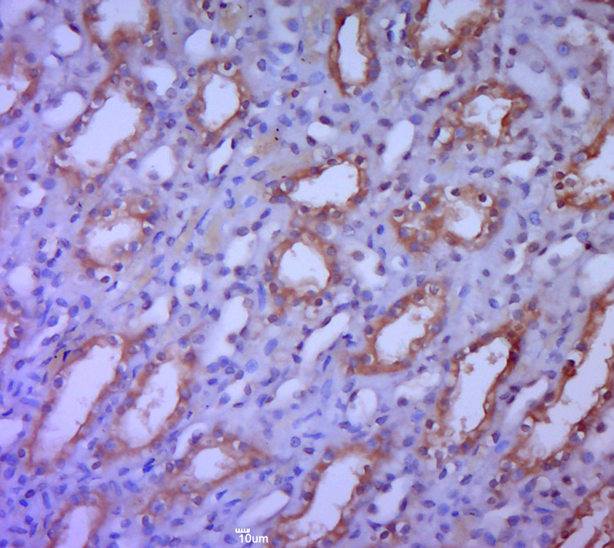
SAV1 Rabbit pAb
SAV1 Rabbit pAb
- 产品详情
- 实验流程
- 背景知识
Application
| IHC-P, IHC-F, IF |
|---|---|
| Primary Accession | Q9H4B6 |
| Reactivity | Rat |
| Predicted | Human, Mouse, Dog, Pig, Horse, Rabbit, Sheep |
| Host | Rabbit |
| Clonality | Polyclonal |
| Calculated MW | 44634 Da |
| Physical State | Liquid |
| Immunogen | KLH conjugated synthetic peptide derived from human SAV1 |
| Epitope Specificity | 1-100/383 |
| Isotype | IgG |
| Purity | affinity purified by Protein A |
| Buffer | 0.01M TBS (pH7.4) with 1% BSA, 0.02% Proclin300 and 50% Glycerol. |
| SUBCELLULAR LOCATION | Nucleus. Cytoplasm. |
| SIMILARITY | Contains 1 SARAH domain. Contains 2 WW domains. |
| Post-translational modifications | Phosphorylated by STK3 and STK4. Phosphorylation is not required for SAV1 stability and may increase the number of protein binding sites on the scaffold molecule. |
| Important Note | This product as supplied is intended for research use only, not for use in human, therapeutic or diagnostic applications. |
| Background Descriptions | WW domain-containing proteins are found in all eukaryotes and play an important role in the regulation of a wide variety of cellular functions such as protein degradation, transcription, and RNA splicing. This gene encodes a protein with two WW domains, a SARAH domain, and a coiled-coil region and is ubiquitously expressed in adult tissues. This protein binds to MST1 (mammalian sterile 20-like kinase 1) and promotes MST1-induced apoptosis. It has also been shown to bind to HAX1 (hematopoietic cell-specific protein 1 (HS1)-associated protein X-1) and to attenuate the anti-apoptotic effects of HAX1. Studies in human and mouse suggest this gene acts as a tumor suppressor. [provided by RefSeq, Aug 2012] |
| Gene ID | 60485 |
|---|---|
| Other Names | Protein salvador homolog 1, 45 kDa WW domain protein, hWW45, SAV1 (HGNC:17795), WW45 |
| Target/Specificity | Ubiquitously expressed in adult tissues with highest expression in the pancreas, aorta and interventricular septum and lowest expression in skeletal muscle. Expression was higher in fetal than in the adult heart. Expressed in various cell lines. |
| Dilution | IHC-P=1:100-500,IHC-F=1:100-500,IF=1:100-500 |
| Storage | Store at -20 °C for one year. Avoid repeated freeze/thaw cycles. When reconstituted in sterile pH 7.4 0.01M PBS or diluent of antibody the antibody is stable for at least two weeks at 2-4 °C. |
| Name | SAV1 (HGNC:17795) |
|---|---|
| Synonyms | WW45 |
| Function | Regulator of STK3/MST2 and STK4/MST1 in the Hippo signaling pathway which plays a pivotal role in organ size control and tumor suppression by restricting proliferation and promoting apoptosis (PubMed:29063833). The core of this pathway is composed of a kinase cascade wherein STK3/MST2 and STK4/MST1, in complex with its regulatory protein SAV1, phosphorylates and activates LATS1/2 in complex with its regulatory protein MOB1, which in turn phosphorylates and inactivates YAP1 oncoprotein and WWTR1/TAZ. Phosphorylation of YAP1 by LATS1/2 inhibits its translocation into the nucleus to regulate cellular genes important for cell proliferation, cell death, and cell migration. SAV1 is required for STK3/MST2 and STK4/MST1 activation and promotes cell- cycle exit and terminal differentiation in developing epithelial tissues. Plays a role in centrosome disjunction by regulating the localization of NEK2 to centrosomes, and its ability to phosphorylate CROCC and CEP250. In conjunction with STK3/MST2, activates the transcriptional activity of ESR1 through the modulation of its phosphorylation. |
| Cellular Location | Nucleus. Cytoplasm |
| Tissue Location | Ubiquitously expressed in adult tissues with highest expression in the pancreas, aorta and interventricular septum and lowest expression in skeletal muscle. Expression was higher in fetal than in the adult heart. Expressed in various cell lines |
For Research Use Only. Not For Use In Diagnostic Procedures.
Provided below are standard protocols that you may find useful for product applications.
BACKGROUND
WW domain-containing proteins are found in all eukaryotes and play an important role in the regulation of a wide variety of cellular functions such as protein degradation, transcription, and RNA splicing. This gene encodes a protein with two WW domains, a SARAH domain, and a coiled-coil region and is ubiquitously expressed in adult tissues. This protein binds to MST1 (mammalian sterile 20-like kinase 1) and promotes MST1-induced apoptosis. It has also been shown to bind to HAX1 (hematopoietic cell-specific protein 1 (HS1)-associated protein X-1) and to attenuate the anti-apoptotic effects of HAX1. Studies in human and mouse suggest this gene acts as a tumor suppressor. [provided by RefSeq, Aug 2012]
终于等到您。ABCEPTA(百远生物)抗体产品。
点击下方“我要评价 ”按钮提交您的反馈信息,您的反馈和评价是我们最宝贵的财富之一,
我们将在1-3个工作日内处理您的反馈信息。
如有疑问,联系:0512-88856768 tech-china@abcepta.com.


