


 癌症的基本特征包括细胞增殖、血管生成、迁移、凋亡逃避机制和细胞永生等。找到癌症发生过程中这些通路的关键标记物和对应的抗体用于检测至关重要。
 为您推荐一个泛素化位点预测神器——泛素化分析工具,可以为您的蛋白的泛素化位点作出预测和评分。
 细胞自噬受体图形绘图工具为你的蛋白的细胞受体结合位点作出预测和评分,识别结合到自噬通路中的蛋白是非常重要的,便于让我们理解自噬在正常生理、病理过程中的作用,如发育、细胞分化、神经退化性疾病、压力条件下、感染和癌症。
HUS1 Rabbit pAb
HUS1 Rabbit pAb
- 产品详情
- 实验流程
- 背景知识
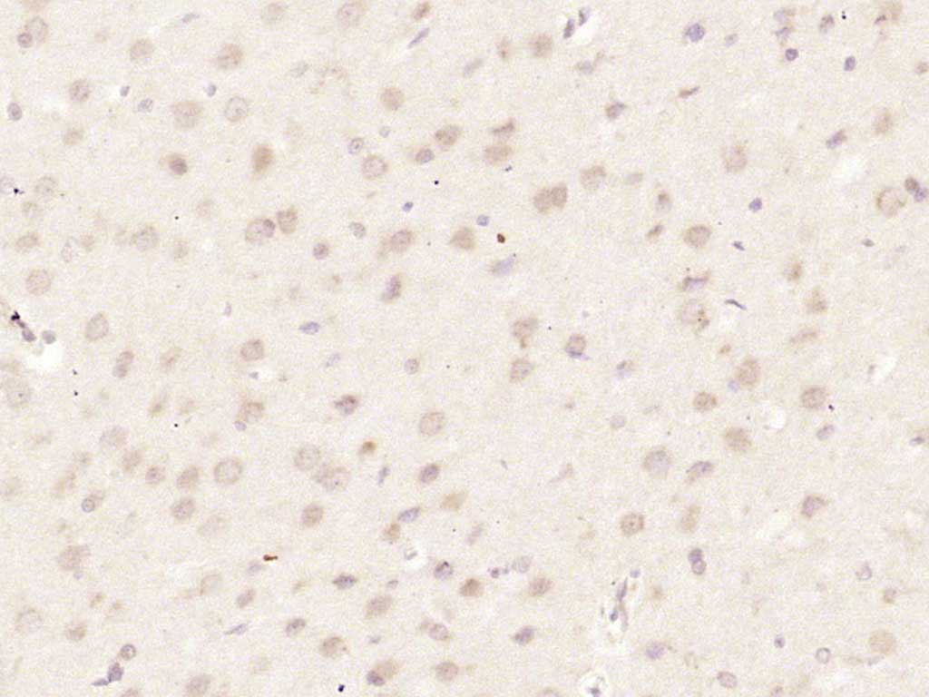
Application
| IHC-P, IHC-F, IF |
|---|---|
| Primary Accession | O60921 |
| Reactivity | Rat |
| Predicted | Human, Mouse, Pig, Horse |
| Host | Rabbit |
| Clonality | Polyclonal |
| Calculated MW | 31691 Da |
| Physical State | Liquid |
| Immunogen | KLH conjugated synthetic peptide derived from human HUS1 |
| Epitope Specificity | 51-150/280 |
| Isotype | IgG |
| Purity | affinity purified by Protein A |
| Buffer | 0.01M TBS (pH7.4) with 1% BSA, 0.02% Proclin300 and 50% Glycerol. |
| SUBCELLULAR LOCATION | Nucleus. Cytoplasm. In discrete nuclear foci upon DNA damage. According to PubMed:14500360, localized also in the cytoplasm. DNA damage induces its nuclear translocation. Shuttles between the nucleus and the cytoplasm. |
| SIMILARITY | Belongs to the HUS1 family. |
| Important Note | This product as supplied is intended for research use only, not for use in human, therapeutic or diagnostic applications. |
| Background Descriptions | The protein encoded by this gene is a component of an evolutionarily conserved, genotoxin-activated checkpoint complex that is involved in the cell cycle arrest in response to DNA damage. This protein forms a heterotrimeric complex with checkpoint proteins RAD9 and RAD1. In response to DNA damage, the trimeric complex interacts with another protein complex consisting of checkpoint protein RAD17 and four small subunits of the replication factor C (RFC), which loads the combined complex onto the chromatin. The DNA damage induced chromatin binding has been shown to depend on the activation of the checkpoint kinase ATM, and is thought to be an early checkpoint signaling event. Alternative splicing results in multiple transcript variants. [provided by RefSeq, Feb 2011] |
| Gene ID | 3364 |
|---|---|
| Other Names | Checkpoint protein HUS1, hHUS1, HUS1 |
| Target/Specificity | Ubiquitous. |
| Dilution | IHC-P=1:100-500,IHC-F=1:100-500,IF=1:100-500 |
| Storage | Store at -20 °C for one year. Avoid repeated freeze/thaw cycles. When reconstituted in sterile pH 7.4 0.01M PBS or diluent of antibody the antibody is stable for at least two weeks at 2-4 °C. |
| Name | HUS1 |
|---|---|
| Function | Component of the 9-1-1 cell-cycle checkpoint response complex that plays a major role in DNA repair (PubMed:21659603). The 9-1-1 complex is recruited to DNA lesion upon damage by the RAD17-replication factor C (RFC) clamp loader complex (PubMed:21659603). Acts then as a sliding clamp platform on DNA for several proteins involved in long- patch base excision repair (LP-BER) (PubMed:21659603). The 9-1-1 complex stimulates DNA polymerase beta (POLB) activity by increasing its affinity for the 3'-OH end of the primer-template and stabilizes POLB to those sites where LP-BER proceeds; endonuclease FEN1 cleavage activity on substrates with double, nick, or gap flaps of distinct sequences and lengths; and DNA ligase I (LIG1) on long-patch base excision repair substrates (PubMed:21659603). The 9-1-1 complex is necessary for the recruitment of RHNO1 to sites of double-stranded breaks (DSB) occurring during the S phase (PubMed:21659603). |
| Cellular Location | Nucleus. Cytoplasm, cytosol. Note=In discrete nuclear foci upon DNA damage (PubMed:11077446). According to PubMed:11077446, localized also in the cytoplasm (PubMed:11077446). DNA damage induces its nuclear translocation (PubMed:11077446). Shuttles between the nucleus and the cytoplasm (PubMed:11077446). |
| Tissue Location | Ubiquitous.. |
For Research Use Only. Not For Use In Diagnostic Procedures.
Provided below are standard protocols that you may find useful for product applications.
BACKGROUND
The protein encoded by this gene is a component of an evolutionarily conserved, genotoxin-activated checkpoint complex that is involved in the cell cycle arrest in response to DNA damage. This protein forms a heterotrimeric complex with checkpoint proteins RAD9 and RAD1. In response to DNA damage, the trimeric complex interacts with another protein complex consisting of checkpoint protein RAD17 and four small subunits of the replication factor C (RFC), which loads the combined complex onto the chromatin. The DNA damage induced chromatin binding has been shown to depend on the activation of the checkpoint kinase ATM, and is thought to be an early checkpoint signaling event. Alternative splicing results in multiple transcript variants. [provided by RefSeq, Feb 2011]
终于等到您。ABCEPTA(百远生物)抗体产品。
点击下方“我要评价 ”按钮提交您的反馈信息,您的反馈和评价是我们最宝贵的财富之一,
我们将在1-3个工作日内处理您的反馈信息。
如有疑问,联系:0512-88856768 tech-china@abcepta.com.


