


 癌症的基本特征包括细胞增殖、血管生成、迁移、凋亡逃避机制和细胞永生等。找到癌症发生过程中这些通路的关键标记物和对应的抗体用于检测至关重要。
 为您推荐一个泛素化位点预测神器——泛素化分析工具,可以为您的蛋白的泛素化位点作出预测和评分。
 细胞自噬受体图形绘图工具为你的蛋白的细胞受体结合位点作出预测和评分,识别结合到自噬通路中的蛋白是非常重要的,便于让我们理解自噬在正常生理、病理过程中的作用,如发育、细胞分化、神经退化性疾病、压力条件下、感染和癌症。
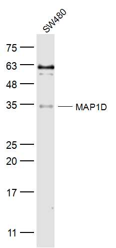
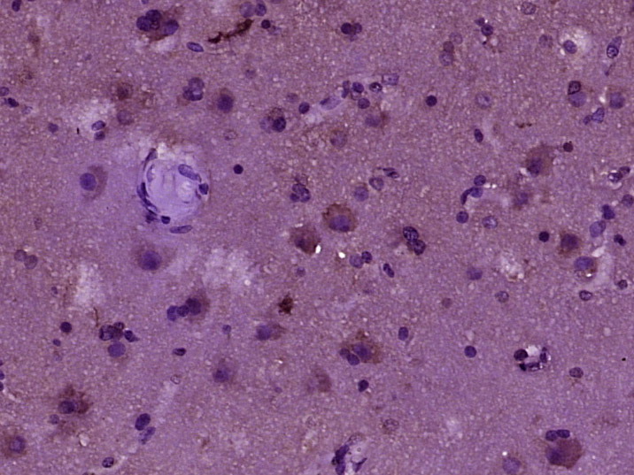
MAP1D Rabbit pAb
MAP1D Rabbit pAb
- 产品详情
- 实验流程
- 背景知识
Application
| WB, IHC-P, IHC-F, IF |
|---|---|
| Primary Accession | Q6UB28 |
| Reactivity | Human |
| Predicted | Mouse, Dog, Horse, Rabbit, Sheep |
| Host | Rabbit |
| Clonality | Polyclonal |
| Calculated MW | 37088 Da |
| Physical State | Liquid |
| Immunogen | KLH conjugated synthetic peptide derived from human MAP1D |
| Epitope Specificity | 251-335/335 |
| Isotype | IgG |
| Purity | affinity purified by Protein A |
| Buffer | 0.01M TBS (pH7.4) with 1% BSA, 0.02% Proclin300 and 50% Glycerol. |
| SUBCELLULAR LOCATION | Mitochondrion. |
| SIMILARITY | Belongs to the peptidase M24A family. |
| Important Note | This product as supplied is intended for research use only, not for use in human, therapeutic or diagnostic applications. |
| Background Descriptions | The N-terminal methionine excision pathway is an essential process in which the N-terminal methionine is removed from many proteins, thus facilitating subsequent protein modification. In mitochondria, enzymes that catalyze this reaction are celled methionine aminopeptidases (MetAps, or MAPs; EC 3.4.11.18) (Serero et al., 2003 [PubMed 14532271]).[supplied by OMIM, Mar 2008] |
| Gene ID | 254042 |
|---|---|
| Other Names | Methionine aminopeptidase 1D, mitochondrial {ECO:0000255|HAMAP-Rule:MF_03174}, MAP 1D {ECO:0000255|HAMAP-Rule:MF_03174}, MetAP 1D {ECO:0000255|HAMAP-Rule:MF_03174}, 3.4.11.18 {ECO:0000255|HAMAP-Rule:MF_03174}, Methionyl aminopeptidase type 1D, mitochondrial, Peptidase M 1D {ECO:0000255|HAMAP-Rule:MF_03174}, METAP1D, MAP1D |
| Target/Specificity | Overexpressed in colon cancer cell lines and colon tumors as compared to normal tissues (at protein level). |
| Dilution | WB=1:500-2000,IHC-P=1:100-500,IHC-F=1:100-500,IF=1:100-500 |
| Storage | Store at -20 °C for one year. Avoid repeated freeze/thaw cycles. When reconstituted in sterile pH 7.4 0.01M PBS or diluent of antibody the antibody is stable for at least two weeks at 2-4 °C. |
| Name | METAP1D |
|---|---|
| Synonyms | MAP1D |
| Function | Removes the N-terminal methionine from nascent proteins. The N-terminal methionine is often cleaved when the second residue in the primary sequence is small and uncharged (Met-Ala-, Cys, Gly, Pro, Ser, Thr, or Val). Requires deformylation of the N(alpha)-formylated initiator methionine before it can be hydrolyzed (By similarity). May play a role in colon tumorigenesis. |
| Cellular Location | Mitochondrion {ECO:0000255|HAMAP-Rule:MF_03174, ECO:0000269|PubMed:14532271} |
| Tissue Location | Overexpressed in colon cancer cell lines and colon tumors as compared to normal tissues (at protein level) |
For Research Use Only. Not For Use In Diagnostic Procedures.
Provided below are standard protocols that you may find useful for product applications.
BACKGROUND
The N-terminal methionine excision pathway is an essential process in which the N-terminal methionine is removed from many proteins, thus facilitating subsequent protein modification. In mitochondria, enzymes that catalyze this reaction are celled methionine aminopeptidases (MetAps, or MAPs; EC 3.4.11.18) (Serero et al., 2003 [PubMed 14532271]).[supplied by OMIM, Mar 2008]
终于等到您。ABCEPTA(百远生物)抗体产品。
点击下方“我要评价 ”按钮提交您的反馈信息,您的反馈和评价是我们最宝贵的财富之一,
我们将在1-3个工作日内处理您的反馈信息。
如有疑问,联系:0512-88856768 tech-china@abcepta.com.


