


 癌症的基本特征包括细胞增殖、血管生成、迁移、凋亡逃避机制和细胞永生等。找到癌症发生过程中这些通路的关键标记物和对应的抗体用于检测至关重要。
 为您推荐一个泛素化位点预测神器——泛素化分析工具,可以为您的蛋白的泛素化位点作出预测和评分。
 细胞自噬受体图形绘图工具为你的蛋白的细胞受体结合位点作出预测和评分,识别结合到自噬通路中的蛋白是非常重要的,便于让我们理解自噬在正常生理、病理过程中的作用,如发育、细胞分化、神经退化性疾病、压力条件下、感染和癌症。
Nance-Horan Syndrome Protein Rabbit pAb
Nance-Horan Syndrome Protein Rabbit pAb
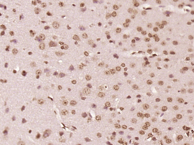
- 产品详情
- 实验流程
- 背景知识
Application 
| IHC-P, IHC-F, IF |
|---|---|
| Primary Accession | Q6T4R5 |
| Reactivity | Rat, Mouse |
| Predicted | Pig, Dog, Human, Rabbit, Chicken, Horse, Sheep |
| Host | Rabbit |
| Clonality | Polyclonal |
| Calculated MW | 179135 Da |
| Physical State | Liquid |
| Immunogen | KLH conjugated synthetic peptide derived from human Nance-Horan Syndrome Protein |
| Epitope Specificity | 1451-1550/1651 |
| Isotype | IgG |
| Purity | affinity purified by Protein A |
| Buffer | 0.01M TBS (pH7.4) with 1% BSA, 0.02% Proclin300 and 50% Glycerol. |
| SUBCELLULAR LOCATION | Nucleus. |
| DISEASE | Nance-Horan syndrome (NHS) [MIM:302350]: Rare X-linked disorder characterized by congenital cataracts, dental anomalies, dysmorphic features, and, in some cases, mental retardation. Distinctive dental anomalies are seen in affected males, including supernumerary incisors and crown shaped permanent teeth. Characteristic facial features are anteverted pinnae, long face, and prominent nasal bridge and nose. Carrier females display milder variable symptoms of disease with lens opacities often involving the posterior Y sutures, and on occasion dental anomalies and the characteristic facial features described. Note: The disease is caused by mutations affecting the gene represented in this entry. Cataract 40 (CTRCT40) [MIM:302200]: An opacification of the crystalline lens of the eye that frequently results in visual impairment or blindness. Opacities vary in morphology, are often confined to a portion of the lens, and may be static or progressive. CTRCT40 manifests as a congenital nuclear opacity with severe visual impairment in affected males. Heterozygous females have suture cataracts and only slight reduction in vision. In some cases, cataract is associated with microcornea without any other systemic anomaly or dysmorphism. Microcornea is defined by a corneal diameter inferior to 10 mm in both meridians in an otherwise normal eye. Note: The disease is caused by mutations affecting the gene represented in this entry. Caused by copy number variations predicted to result in altered transcriptional regulation of the NHS gene: a 0.8 Mb segmental duplication-triplication encompassing the NHS, SCML1 and RAI2 genes, and an 4.8 kb intragenic deletion in NHS intron 1. |
| Important Note | This product as supplied is intended for research use only, not for use in human, therapeutic or diagnostic applications. |
| Background Descriptions | This gene encodes a protein containing four conserved nuclear localization signals. The encoded protein functions in eye, tooth, craniofacial and brain development, and it can regulate actin remodeling and cell morphology. Mutations in this gene have been shown to cause Nance-Horan syndrome, and also X-linked cataract-40. Alternatively spliced transcript variants encoding different isoforms have been described for this gene. [provided by RefSeq, May 2014] |
| Gene ID | 4810 |
|---|---|
| Other Names | Actin remodeling regulator NHS, Congenital cataracts and dental anomalies protein, Nance-Horan syndrome protein, NHS |
| Dilution | IHC-P=1:100-500,IHC-F=1:100-500,IF=1:100-500 |
| Storage | Store at -20 °C for one year. Avoid repeated freeze/thaw cycles. When reconstituted in sterile pH 7.4 0.01M PBS or diluent of antibody the antibody is stable for at least two weeks at 2-4 °C. |
For Research Use Only. Not For Use In Diagnostic Procedures.
| Name | NHS |
|---|---|
| Function | May function in cell morphology by maintaining the integrity of the circumferential actin ring and controlling lamellipod formation. Involved in the regulation eye, tooth, brain and craniofacial development. |
| Cellular Location | [Isoform 1]: Apical cell membrane; Peripheral membrane protein. Cell projection, lamellipodium. Cell junction, tight junction. Cell junction, focal adhesion. Note=Colocalizes with the tight junction protein TJP1 in epithelial cells. Localizes to the leading edge of lamellipodia in motile cells |
| Tissue Location | Detected at low levels in all tissues analyzed. Detected in fetal and adult brain, lens, retina, retinal pigment epithelium, placenta, lymphocytes and fibroblasts. Levels in retinal pigment epithelium, placenta, lymphocytes, and fibroblasts are very low. Expressed also in kidney, lung and thymus |
Research Areas
Application Protocols
Provided below are standard protocols that you may find useful for product applications.
BACKGROUND
This product as supplied is intended for research use only, not for use in human, therapeutic or diagnostic applications.

终于等到您。ABCEPTA(百远生物)抗体产品。
点击下方“我要评价 ”按钮提交您的反馈信息,您的反馈和评价是我们最宝贵的财富之一,
我们将在1-3个工作日内处理您的反馈信息。
如有疑问,联系:0512-88856768 tech-china@abcepta.com.


