


 癌症的基本特征包括细胞增殖、血管生成、迁移、凋亡逃避机制和细胞永生等。找到癌症发生过程中这些通路的关键标记物和对应的抗体用于检测至关重要。
 为您推荐一个泛素化位点预测神器——泛素化分析工具,可以为您的蛋白的泛素化位点作出预测和评分。
 细胞自噬受体图形绘图工具为你的蛋白的细胞受体结合位点作出预测和评分,识别结合到自噬通路中的蛋白是非常重要的,便于让我们理解自噬在正常生理、病理过程中的作用,如发育、细胞分化、神经退化性疾病、压力条件下、感染和癌症。
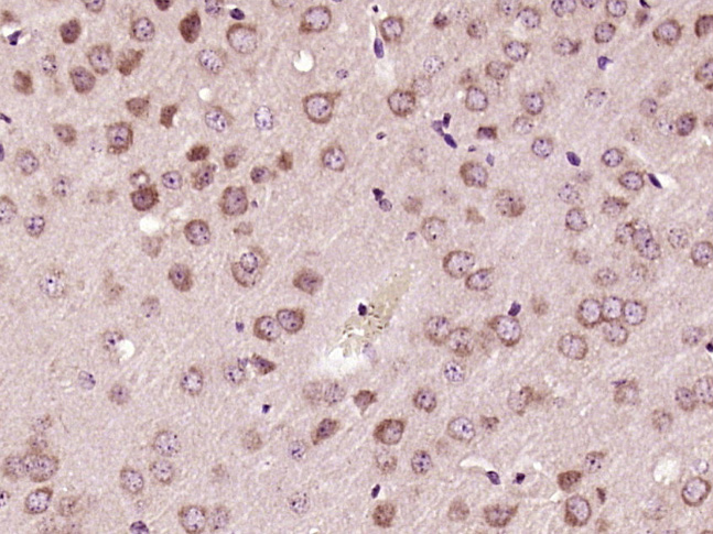
SH3BP1 Rabbit pAb
SH3BP1 Rabbit pAb
- 产品详情
- 实验流程
Application
| IHC-P, IHC-F, IF |
|---|---|
| Primary Accession | Q9Y3L3 |
| Reactivity | Mouse |
| Predicted | Human, Rat, Dog, Pig, Horse, Rabbit |
| Host | Rabbit |
| Clonality | Polyclonal |
| Calculated MW | 75713 Da |
| Physical State | Liquid |
| Immunogen | KLH conjugated synthetic peptide derived from human SH3BP1 |
| Epitope Specificity | 101-200/701 |
| Isotype | IgG |
| Purity | affinity purified by Protein A |
| Buffer | 0.01M TBS (pH7.4) with 1% BSA, 0.02% Proclin300 and 50% Glycerol. |
| SIMILARITY | Contains 1 BAR domain. Contains 1 Rho-GAP domain. |
| Important Note | This product as supplied is intended for research use only, not for use in human, therapeutic or diagnostic applications. |
| Gene ID | 23616 |
|---|---|
| Other Names | SH3 domain-binding protein 1 {ECO:0000312|HGNC:HGNC:10824}, SH3BP1 (HGNC:10824) |
| Dilution | IHC-P=1:100-500,IHC-F=1:100-500,IF=1:100-500 |
| Storage | Store at -20 °C for one year. Avoid repeated freeze/thaw cycles. When reconstituted in sterile pH 7.4 0.01M PBS or diluent of antibody the antibody is stable for at least two weeks at 2-4 °C. |
| Name | SH3BP1 (HGNC:10824) |
|---|---|
| Function | GTPase activating protein (GAP) which specifically converts GTP-bound Rho-type GTPases including RAC1 and CDC42 in their inactive GDP-bound form. By specifically inactivating RAC1 at the leading edge of migrating cells, it regulates the spatiotemporal organization of cell protrusions which is important for proper cell migration (PubMed:21658605). Also negatively regulates CDC42 in the process of actin remodeling and the formation of epithelial cell junctions (PubMed:22891260). Through its GAP activity toward RAC1 and/or CDC42 plays a specific role in phagocytosis of large particles. Specifically recruited by a PI3 kinase/PI3K-dependent mechanism to sites of large particles engagement, inactivates RAC1 and/or CDC42 allowing the reorganization of the underlying actin cytoskeleton required for engulfment (PubMed:26465210). It also plays a role in angiogenesis and the process of repulsive guidance as part of a semaphorin-plexin signaling pathway. Following the binding of PLXND1 to extracellular SEMA3E it dissociates from PLXND1 and inactivates RAC1, inducing the intracellular reorganization of the actin cytoskeleton and the collapse of cells (PubMed:24841563). |
| Cellular Location | Cell projection. Cell junction, tight junction. Cell junction, adherens junction. Cell projection, phagocytic cup. Nucleus Cytoplasm, cytosol. Note=Localizes at the leading edge of migrating cells (PubMed:21658605, PubMed:24841563) Accumulation at forming phagocytic cups is PI3 kinase/PI3K-dependent and is specific for sites of large particles engagement and their phosphatidylinositol 3,4,5-triphosphate membrane content (PubMed:26465210). |
Research Areas
For Research Use Only. Not For Use In Diagnostic Procedures.
Application Protocols
Provided below are standard protocols that you may find useful for product applications.
终于等到您。ABCEPTA(百远生物)抗体产品。
点击下方“我要评价 ”按钮提交您的反馈信息,您的反馈和评价是我们最宝贵的财富之一,
我们将在1-3个工作日内处理您的反馈信息。
如有疑问,联系:0512-88856768 tech-china@abcepta.com.


