


 癌症的基本特征包括细胞增殖、血管生成、迁移、凋亡逃避机制和细胞永生等。找到癌症发生过程中这些通路的关键标记物和对应的抗体用于检测至关重要。
 为您推荐一个泛素化位点预测神器——泛素化分析工具,可以为您的蛋白的泛素化位点作出预测和评分。
 细胞自噬受体图形绘图工具为你的蛋白的细胞受体结合位点作出预测和评分,识别结合到自噬通路中的蛋白是非常重要的,便于让我们理解自噬在正常生理、病理过程中的作用,如发育、细胞分化、神经退化性疾病、压力条件下、感染和癌症。
CD5L Rabbit pAb
CD5L Rabbit pAb
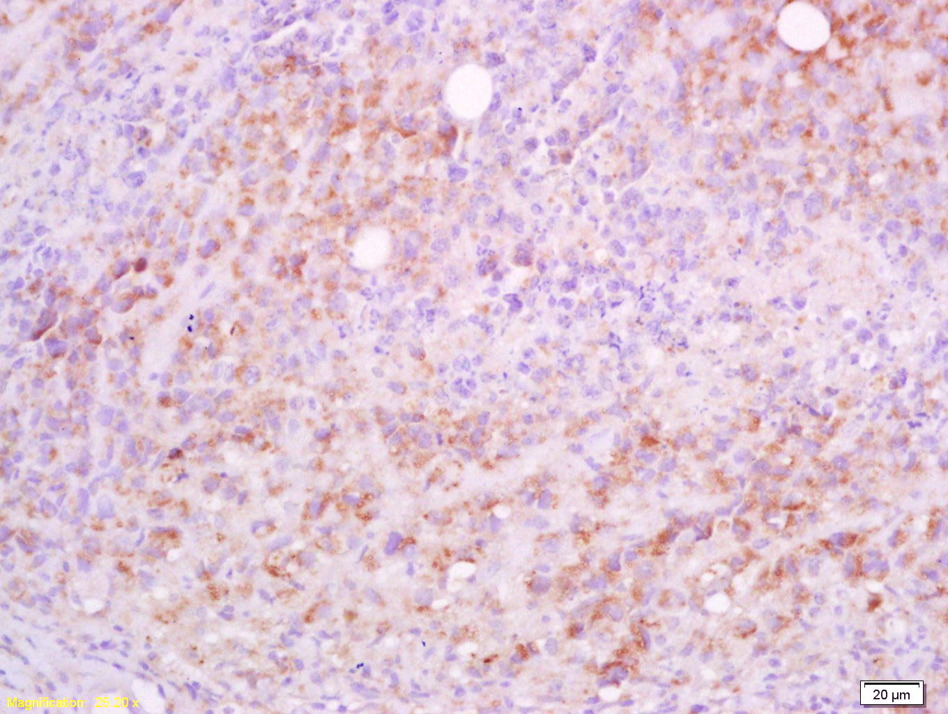
- 产品详情
- 实验流程
- 背景知识
Application 
| IHC-P, IHC-F, IF |
|---|---|
| Primary Accession | O43866 |
| Reactivity | Rat, Mouse |
| Predicted | Human |
| Host | Rabbit |
| Clonality | Polyclonal |
| Calculated MW | 38088 Da |
| Physical State | Liquid |
| Immunogen | KLH conjugated synthetic peptide derived from human CD5L |
| Epitope Specificity | 221-347/347 |
| Isotype | IgG |
| Purity | affinity purified by Protein A |
| Buffer | 0.01M TBS (pH7.4) with 1% BSA, 0.02% Proclin300 and 50% Glycerol. |
| SUBCELLULAR LOCATION | Secreted. |
| Important Note | This product as supplied is intended for research use only, not for use in human, therapeutic or diagnostic applications. |
| Background Descriptions | CD5L is a member of the scavenger receptor cysteine-rich domain superfamily (SRCR-SF) initially identified as an inducible cell surface ligand of CD5. It was shown that CD5L functions in the thymus as the inducer of resistance to apoptosis within CD4+/CD8+ thymocytes and as the supporter of the viability of these cells before thymic selection. CD5L was also shown to support macrophage survival and enhance their phagocytic function. More recent experiments using recombinant CD5L significantly inhibited apoptosis of NKT and T cells obtained from C. parvum-stimulated livers in vitro, suggesting that CD5L functions to induce resistance to apoptosis in these cells and supports host defense against inflammation during infection. Expressed in spleen, lymph node, thymus, bone marrow, and fetal liver, but not in non-lymphoid tissues. |
| Gene ID | 922 |
|---|---|
| Other Names | CD5 antigen-like, Apoptosis inhibitor expressed by macrophages, hAIM, CT-2 {ECO:0000303|Ref.2}, IgM-associated peptide, SP-alpha, CD5L, API6 |
| Dilution | IHC-P=1:100-500,IHC-F=1:100-500,IF=1:100-500 |
| Storage | Store at -20 °C for one year. Avoid repeated freeze/thaw cycles. When reconstituted in sterile pH 7.4 0.01M PBS or diluent of antibody the antibody is stable for at least two weeks at 2-4 °C. |
For Research Use Only. Not For Use In Diagnostic Procedures.
| Name | CD5L |
|---|---|
| Synonyms | API6 |
| Function | Secreted protein that acts as a key regulator of lipid synthesis: mainly expressed by macrophages in lymphoid and inflamed tissues and regulates mechanisms in inflammatory responses, such as infection or atherosclerosis. Able to inhibit lipid droplet size in adipocytes. Following incorporation into mature adipocytes via CD36- mediated endocytosis, associates with cytosolic FASN, inhibiting fatty acid synthase activity and leading to lipolysis, the degradation of triacylglycerols into glycerol and free fatty acids (FFA). CD5L-induced lipolysis occurs with progression of obesity: participates in obesity- associated inflammation following recruitment of inflammatory macrophages into adipose tissues, a cause of insulin resistance and obesity-related metabolic disease. Regulation of intracellular lipids mediated by CD5L has a direct effect on transcription regulation mediated by nuclear receptors ROR-gamma (RORC). Acts as a key regulator of metabolic switch in T-helper Th17 cells. Regulates the expression of pro-inflammatory genes in Th17 cells by altering the lipid content and limiting synthesis of cholesterol ligand of RORC, the master transcription factor of Th17-cell differentiation. CD5L is mainly present in non-pathogenic Th17 cells, where it decreases the content of polyunsaturated fatty acyls (PUFA), affecting two metabolic proteins MSMO1 and CYP51A1, which synthesize ligands of RORC, limiting RORC activity and expression of pro-inflammatory genes. Participates in obesity-associated autoimmunity via its association with IgM, interfering with the binding of IgM to Fcalpha/mu receptor and enhancing the development of long-lived plasma cells that produce high- affinity IgG autoantibodies (By similarity). Also acts as an inhibitor of apoptosis in macrophages: promotes macrophage survival from the apoptotic effects of oxidized lipids in case of atherosclerosis (PubMed:24295828). Involved in early response to microbial infection against various pathogens by acting as a pattern recognition receptor and by promoting autophagy (PubMed:16030018, PubMed:24223991, PubMed:24583716, PubMed:25713983). Promotes recovery from acute kidney injury (AKI) by accumulating on necrotic cell debris within the kidney proximal tubules following AKI and interacting with HAVCR1 expressed on the surface of injured kidney tubular epithelial cells (PubMed:26726878). This enhances HAVCR1-mediated phagocytosis of intraluminal necrotic debris and contributes to kidney tissue repair (PubMed:26726878). Also enhances necrotic debris uptake by kidney macrophages in a HAVCR1-independent manner (By similarity). |
| Cellular Location | Secreted. Cytoplasm {ECO:0000250|UniProtKB:Q9QWK4}. Note=Secreted by macrophages and circulates in the blood (PubMed:24223991, PubMed:24804991). Transported into the cytoplasm via CD36-mediated endocytosis (By similarity) Following acute kidney injury, dissociates from IgM in the blood and is excreted into urine, leading to its accumulation at intraluminal debris in the kidney proximal tubules (PubMed:26726878) {ECO:0000250|UniProtKB:Q9QWK4, ECO:0000269|PubMed:24223991, ECO:0000269|PubMed:24804991, ECO:0000269|PubMed:26726878} |
| Tissue Location | Expressed in spleen, lymph node, thymus, bone marrow, and fetal liver, but not in non-lymphoid tissues |
Research Areas
Application Protocols
Provided below are standard protocols that you may find useful for product applications.
BACKGROUND
This product as supplied is intended for research use only, not for use in human, therapeutic or diagnostic applications.

终于等到您。ABCEPTA(百远生物)抗体产品。
点击下方“我要评价 ”按钮提交您的反馈信息,您的反馈和评价是我们最宝贵的财富之一,
我们将在1-3个工作日内处理您的反馈信息。
如有疑问,联系:0512-88856768 tech-china@abcepta.com.


