


 癌症的基本特征包括细胞增殖、血管生成、迁移、凋亡逃避机制和细胞永生等。找到癌症发生过程中这些通路的关键标记物和对应的抗体用于检测至关重要。
 为您推荐一个泛素化位点预测神器——泛素化分析工具,可以为您的蛋白的泛素化位点作出预测和评分。
 细胞自噬受体图形绘图工具为你的蛋白的细胞受体结合位点作出预测和评分,识别结合到自噬通路中的蛋白是非常重要的,便于让我们理解自噬在正常生理、病理过程中的作用,如发育、细胞分化、神经退化性疾病、压力条件下、感染和癌症。
HEPACAM Polyclonal Antibody
Purified Rabbit Polyclonal Antibody (Pab)
- 产品详情
- 实验流程
Application
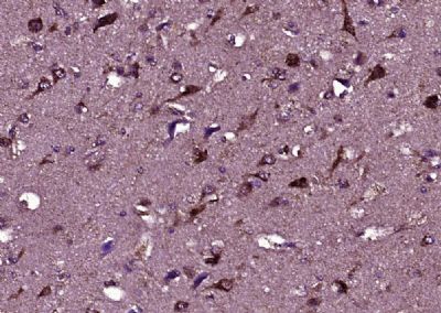
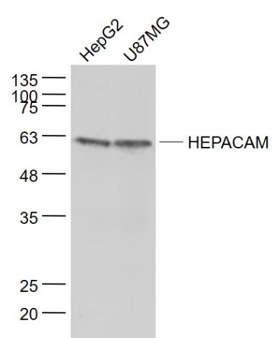
| WB, IHC-P, IHC-F, IF, E |
|---|---|
| Primary Accession | Q14CZ8 |
| Reactivity | Rat, Pig, Dog, Bovine |
| Host | Rabbit |
| Clonality | Polyclonal |
| Calculated MW | 46026 Da |
| Physical State | Liquid |
| Immunogen | KLH conjugated synthetic peptide derived from human HEPACAM |
| Epitope Specificity | 101-200/416 |
| Isotype | IgG |
| Purity | affinity purified by Protein A |
| Buffer | 0.01M TBS (pH7.4) with 1% BSA, 0.02% Proclin300 and 50% Glycerol. |
| SUBCELLULAR LOCATION | Cytoplasm. Membrane; Single-pass type I membrane protein; Cytoplasmic side. Note=In MCF7 breast carcinoma and hepatic Hep3B and HepG2 cell lines, localization of HEPACAM is cell density-dependent. In well spread cells, localized to punctate structures in the perinuclear membrane, cytoplasm, and at cell surface of protusions. In confluent cells, localized predominantly to the cytoplasmic membrane, particularly in areas of cell-cell contacts. Colocalizes with CDH1. |
| SIMILARITY | Contains 1 Ig-like C2-type (immunoglobulin-like) domain.Contains 1 Ig-like V-type (immunoglobulin-like) domain. |
| SUBUNIT | Homodimer. Dimer formation occurs predominantly through cis interactions on the cell surface. |
| Important Note | This product as supplied is intended for research use only, not for use in human, therapeutic or diagnostic applications. |
| Background Descriptions | Involved in regulating cell motility and cell-matrix interactions. May inhibit cell growth through suppression of cell proliferation. |
| Gene ID | 220296 |
|---|---|
| Other Names | Hepatocyte cell adhesion molecule, Protein hepaCAM, HEPACAM {ECO:0000312|EMBL:AAI13563.1} |
| Dilution | WB=1:500-2000,IHC-P=1:100-500,IHC-F=1:100-500,IF=1:100-500,ELISA=1:5000-10000 |
| Format | 0.01M TBS(pH7.4) with 1% BSA, 0.09% (W/V) sodium azide and 50% Glyce |
| Storage | Store at -20 °C for one year. Avoid repeated freeze/thaw cycles. When reconstituted in sterile pH 7.4 0.01M PBS or diluent of antibody the antibody is stable for at least two weeks at 2-4 °C. |
| Name | HEPACAM {ECO:0000303|PubMed:15885354, ECO:0000312|HGNC:HGNC:26361} |
|---|---|
| Function | Involved in regulating cell motility and cell-matrix interactions. May inhibit cell growth through suppression of cell proliferation (PubMed:15885354, PubMed:15917256). In glia, associates and targets CLCN2 at astrocytic processes and myelinated fiber tracts where it may regulate transcellular chloride flux involved in neuron excitability (PubMed:22405205). |
| Cellular Location | Cytoplasm. Cell membrane; Single-pass type I membrane protein; Cytoplasmic side. Note=Colocalizes with CLCN2 at astrocyte end-foot in contact with brain capillaries and other glial cells (By similarity). In MCF-7 breast carcinoma and hepatic Hep 3B2.1-7 and Hep- G2 cell lines, localization of HEPACAM is cell density-dependent. In well spread cells, localized to punctate structures in the perinuclear membrane, cytoplasm, and at cell surface of protusions. In confluent cells, localized predominantly to the cytoplasmic membrane, particularly in areas of cell-cell contacts. Colocalizes with CDH1 {ECO:0000250|UniProtKB:Q640R3} |
Research Areas
For Research Use Only. Not For Use In Diagnostic Procedures.
Application Protocols
Provided below are standard protocols that you may find useful for product applications.
终于等到您。ABCEPTA(百远生物)抗体产品。
点击下方“我要评价 ”按钮提交您的反馈信息,您的反馈和评价是我们最宝贵的财富之一,
我们将在1-3个工作日内处理您的反馈信息。
如有疑问,联系:0512-88856768 tech-china@abcepta.com.


