


 癌症的基本特征包括细胞增殖、血管生成、迁移、凋亡逃避机制和细胞永生等。找到癌症发生过程中这些通路的关键标记物和对应的抗体用于检测至关重要。
 为您推荐一个泛素化位点预测神器——泛素化分析工具,可以为您的蛋白的泛素化位点作出预测和评分。
 细胞自噬受体图形绘图工具为你的蛋白的细胞受体结合位点作出预测和评分,识别结合到自噬通路中的蛋白是非常重要的,便于让我们理解自噬在正常生理、病理过程中的作用,如发育、细胞分化、神经退化性疾病、压力条件下、感染和癌症。
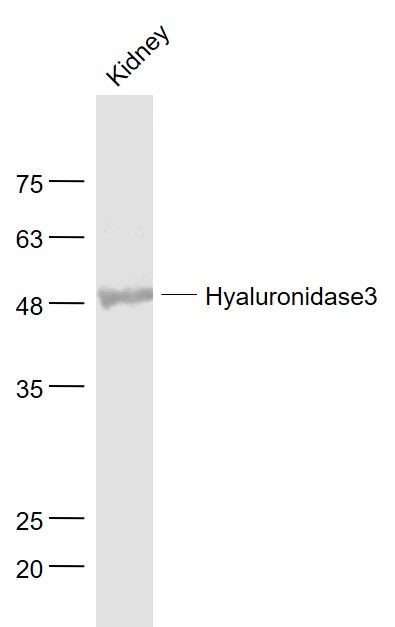
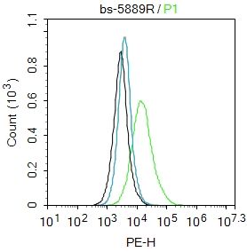
Hyaluronidase3 Polyclonal Antibody
Purified Rabbit Polyclonal Antibody (Pab)
- 产品详情
- 实验流程
Application
| WB, IHC-P, IHC-F, IF, E |
|---|---|
| Primary Accession | O43820 |
| Reactivity | Rat, Pig |
| Host | Rabbit |
| Clonality | Polyclonal |
| Calculated MW | 46501 Da |
| Physical State | Liquid |
| Immunogen | KLH conjugated synthetic peptide derived from human Hyaluronidase3 |
| Epitope Specificity | 51-150/417 |
| Isotype | IgG |
| Purity | affinity purified by Protein A |
| Buffer | 0.01M TBS (pH7.4) with 1% BSA, 0.02% Proclin300 and 50% Glycerol. |
| SUBCELLULAR LOCATION | Secreted (By similarity). Lysosome (By similarity). |
| SIMILARITY | Belongs to the glycosyl hydrolase 56 family. Contains 1 EGF-like domain. |
| Important Note | This product as supplied is intended for research use only, not for use in human, therapeutic or diagnostic applications. |
| Background Descriptions | HYAL3 is a protein which is similar in structure to hyaluronidases. Hyaluronidases intracellularly degrade hyaluronan, one of the major glycosaminoglycans of the extracellular matrix. Hyaluronan is thought to be involved in cell proliferation, migration and differentiation. However, this protein has not yet been shown to have hyaluronidase activity. |
| Gene ID | 8372 |
|---|---|
| Other Names | Hyaluronidase-3, Hyal-3, 3.2.1.35, Hyaluronoglucosaminidase-3, Lung carcinoma protein 3, LuCa-3, HYAL3, LUCA3 |
| Target/Specificity | Bone marrow, testis and kidney. Isoform 4 is detected in all bladder tumor and prostate tumor cells. |
| Dilution | WB=1:500-2000,IHC-P=1:100-500,IHC-F=1:100-500,IF=1:100-500,Flow-Cyt=2ug/Test,ELISA=1:5000-10000 |
| Format | 0.01M TBS(pH7.4) with 1% BSA, 0.09% (W/V) sodium azide and 50% Glyce |
| Storage | Store at -20 °C for one year. Avoid repeated freeze/thaw cycles. When reconstituted in sterile pH 7.4 0.01M PBS or diluent of antibody the antibody is stable for at least two weeks at 2-4 °C. |
| Name | HYAL3 |
|---|---|
| Synonyms | LUCA3 |
| Function | Facilitates sperm penetration into the layer of cumulus cells surrounding the egg by digesting hyaluronic acid. Involved in induction of the acrosome reaction in the sperm. Involved in follicular atresia, the breakdown of immature ovarian follicles that are not selected to ovulate. Induces ovarian granulosa cell apoptosis, possibly via apoptotic signaling pathway involving CASP8 and CASP3 activation, and poly(ADP-ribose) polymerase (PARP) cleavage. Has no hyaluronidase activity in embryonic fibroblasts in vitro. Has no hyaluronidase activity in granulosa cells in vitro. |
| Cellular Location | Secreted {ECO:0000250|UniProtKB:Q8VEI3}. Cell membrane {ECO:0000250|UniProtKB:Q8VEI3}. Cytoplasmic vesicle, secretory vesicle, acrosome {ECO:0000250|UniProtKB:Q8VEI3}. Endoplasmic reticulum {ECO:0000250|UniProtKB:Q8VEI3}. Early endosome {ECO:0000250|UniProtKB:Q8VEI3}. Note=Mostly present in low-density vesicles. Low levels in higher density vesicles of late endosomes and lysosomes. Localized in punctate cytoplasmic vesicles and in perinuclear structures, but does not colocalize with LAMP1. Localized on the plasma membrane over the acrosome and on the surface of the midpiece of the sperm tail. {ECO:0000250|UniProtKB:Q8VEI3} |
| Tissue Location | Expressed in sperm (PubMed:20586096). Highly expressed in epidermis of the skin, where it is expressed intracellularily in the deep horny layer (at protein level) (PubMed:21699545). Bone marrow, testis and kidney (PubMed:10493834) |
Research Areas
For Research Use Only. Not For Use In Diagnostic Procedures.
Application Protocols
Provided below are standard protocols that you may find useful for product applications.
终于等到您。ABCEPTA(百远生物)抗体产品。
点击下方“我要评价 ”按钮提交您的反馈信息,您的反馈和评价是我们最宝贵的财富之一,
我们将在1-3个工作日内处理您的反馈信息。
如有疑问,联系:0512-88856768 tech-china@abcepta.com.
¥ 1,500.00
Cat# AP58341


