


 癌症的基本特征包括细胞增殖、血管生成、迁移、凋亡逃避机制和细胞永生等。找到癌症发生过程中这些通路的关键标记物和对应的抗体用于检测至关重要。
 为您推荐一个泛素化位点预测神器——泛素化分析工具,可以为您的蛋白的泛素化位点作出预测和评分。
 细胞自噬受体图形绘图工具为你的蛋白的细胞受体结合位点作出预测和评分,识别结合到自噬通路中的蛋白是非常重要的,便于让我们理解自噬在正常生理、病理过程中的作用,如发育、细胞分化、神经退化性疾病、压力条件下、感染和癌症。
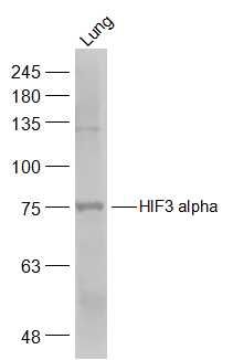
HIF3 alpha Polyclonal Antibody
Purified Rabbit Polyclonal Antibody (Pab)
- 产品详情
- 实验流程
Application
| WB, IHC-P, IHC-F, IF, ICC, E |
|---|---|
| Primary Accession | Q9Y2N7 |
| Reactivity | Human, Mouse, Rat |
| Predicted | Pig, Dog, Horse |
| Host | Rabbit |
| Clonality | Polyclonal |
| Calculated MW | 72433 Da |
| Physical State | Liquid |
| Immunogen | KLH conjugated synthetic peptide derived from human HIF3 alpha |
| Epitope Specificity | 131-230/669 |
| Isotype | IgG |
| Purity | affinity purified by Protein A |
| Buffer | 0.01M TBS (pH7.4) with 1% BSA, 0.02% Proclin300 and 50% Glycerol. |
| SUBCELLULAR LOCATION | Nucleus. Cytoplasm. Note=In the nuclei of all periportal and perivenous hepatocytes. In the distal perivenous zone, detected in the cytoplasm of the hepatocytes. |
| SIMILARITY | Contains 1 basic helix-loop-helix (bHLH) domain.Contains 2 PAS (PER-ARNT-SIM) domains. |
| SUBUNIT | Heterodimerizes with ARNT. Interacts via the oxygen-dependent degradation domain (ODD) with the beta domain of VHL. |
| Important Note | This product as supplied is intended for research use only, not for use in human, therapeutic or diagnostic applications. |
| Background Descriptions | Hypoxia-inducible factor (HIF) is one of the most important factors in the cellular response to hypoxia, transcriptionally activating genes encoding proteins that mediate adaptive responses to reduced oxygen availability. HIF is a heterodimer consisting of one of three subunits, HIF1 alpha, HIF2 alpha, or HIF3 alpha. HIF target genes play critical roles in metabolism, angiogenesis, cell proliferation and cell survival. HIF3 alpha protein is one of several alpha/beta-subunit heterodimeric transcription factors that regulate many adaptive responses to low oxygen tension (hypoxia). The alpha 3 subunit lacks the transactivation domain found in factors containing either the alpha 1 or alpha 2 subunits. HIF3 alpha may be a marker for tumor growth and angiogenesis. |
| Gene ID | 64344 |
|---|---|
| Other Names | Hypoxia-inducible factor 3-alpha, HIF-3-alpha, HIF3-alpha, Basic-helix-loop-helix-PAS protein MOP7, Class E basic helix-loop-helix protein 17, bHLHe17, HIF3-alpha-1, Inhibitory PAS domain protein, IPAS, Member of PAS protein 7, PAS domain-containing protein 7, HIF3A (HGNC:15825), BHLHE17, MOP7, PASD7 |
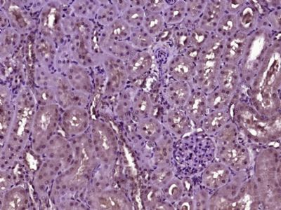
| Target/Specificity | Expressed in kidney. Expressed abundantly in lung epithelial cells. Expression is regulated in an oxygen-dependent manner. |
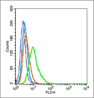
| Dilution | WB=1:500-2000,IHC-P=1:100-500,IHC-F=1:100-500,ICC=1:50,IF=1:100-500,Flow-Cyt=1 µg/Test,ELISA=1:5000-10000 |
| Format | 0.01M TBS(pH7.4) with 1% BSA, 0.09% (W/V) sodium azide and 50% Glyce |
| Storage | Store at -20 °C for one year. Avoid repeated freeze/thaw cycles. When reconstituted in sterile pH 7.4 0.01M PBS or diluent of antibody the antibody is stable for at least two weeks at 2-4 °C. |
| Name | HIF3A (HGNC:15825) |
|---|---|
| Synonyms | BHLHE17, MOP7, PASD7 |
| Function | Acts as a transcriptional regulator in adaptive response to low oxygen tension. Acts as a regulator of hypoxia-inducible gene expression (PubMed:11573933, PubMed:16126907, PubMed:19694616, PubMed:20416395, PubMed:21069422). Functions as an inhibitor of angiogenesis in hypoxic cells of the cornea. Plays a role in the development of the cardiorespiratory system. May also be involved in apoptosis (By similarity). |
| Cellular Location | Nucleus. Cytoplasm Nucleus speckle {ECO:0000250|UniProtKB:Q0VBL6}. Mitochondrion {ECO:0000250|UniProtKB:Q0VBL6}. Note=In the nuclei of all periportal and perivenous hepatocytes. In the distal perivenous zone, detected in the cytoplasm of the hepatocytes. Shuttles between the nucleus and the cytoplasm in a CRM1-dependent manner. Colocalizes with BAD in the cytoplasm. Colocalizes with EPAS1 and HIF1A in the nucleus and speckles (By similarity). Localized in the cytoplasm and nuclei under normoxia, but increased in the nucleus under hypoxic conditions (PubMed:19694616). Colocalized with HIF1A in kidney tumors (PubMed:19694616). {ECO:0000250|UniProtKB:Q0VBL6, ECO:0000250|UniProtKB:Q9JHS2, ECO:0000269|PubMed:19694616} |
| Tissue Location | Expressed in vascular cells (at protein level) (PubMed:21069422). Expressed in kidney (PubMed:11573933, PubMed:19694616). Expressed in lung epithelial cells (PubMed:16775626) Expressed in endothelial cells (venous and arterial cells from umbilical cord and aortic endothelial cells) and in vascular smooth muscle cells (aorta) (PubMed:21069422). Strongly expressed in the heart, placenta, and skeletal muscle, whereas a weak expression profile was found in the lung, liver, and kidney (PubMed:12538644). Expressed weakly in cell renal cell carcinoma (CC-RCC) compared to normal renal cells (PubMed:16126907). Expression is down-regulated in numerous kidney tumor cells compared to non tumor kidney tissues (PubMed:16126907). Isoform 2 is expressed in heart, placenta, lung, liver, skeletal muscle and pancreas and in numerous cancer cell lines (PubMed:20416395). Isoform 3 and isoform 4 are weakly expressed in heart, placenta, lung, liver, skeletal muscle and pancreas (PubMed:20416395). Isoform 4 is expressed in fetal tissues, such as heart, brain, thymus, lung, liver, skeletal kidney and spleen (PubMed:20416395). Isoform 3 is weakly expressed in fetal tissues, such as liver and kidney (PubMed:20416395). |
For Research Use Only. Not For Use In Diagnostic Procedures.
Application Protocols
Provided below are standard protocols that you may find useful for product applications.
终于等到您。ABCEPTA(百远生物)抗体产品。
点击下方“我要评价 ”按钮提交您的反馈信息,您的反馈和评价是我们最宝贵的财富之一,
我们将在1-3个工作日内处理您的反馈信息。
如有疑问,联系:0512-88856768 tech-china@abcepta.com.


