


 癌症的基本特征包括细胞增殖、血管生成、迁移、凋亡逃避机制和细胞永生等。找到癌症发生过程中这些通路的关键标记物和对应的抗体用于检测至关重要。
 为您推荐一个泛素化位点预测神器——泛素化分析工具,可以为您的蛋白的泛素化位点作出预测和评分。
 细胞自噬受体图形绘图工具为你的蛋白的细胞受体结合位点作出预测和评分,识别结合到自噬通路中的蛋白是非常重要的,便于让我们理解自噬在正常生理、病理过程中的作用,如发育、细胞分化、神经退化性疾病、压力条件下、感染和癌症。
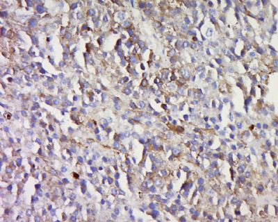
Melan A Rabbit pAb
Melan A Rabbit pAb

- 产品详情
- 实验流程
- 背景知识
Application
| WB |
|---|---|
| Reactivity | Human |
| Host | Rabbit |
| Clonality | Polyclonal |
| Calculated MW | 13 KDa |
| Physical State | Liquid |
| Immunogen | KLH conjugated synthetic peptide derived from mouse Melan A |
| Epitope Specificity | 1-80/113 |
| Isotype | IgG |
| Purity | affinity purified by Protein A |
| Buffer | 0.01M TBS (pH7.4) with 1% BSA, 0.02% Proclin300 and 50% Glycerol. |
| SUBCELLULAR LOCATION | Endoplasmic reticulum membrane; Single-pass type III membrane protein. Golgi apparatus. Golgi apparatus, trans-Golgi network membrane. Melanosome. Note=Also found in small vesicles and tubules dispersed over the entire cytoplasm. A small fraction of the protein is inserted into the membrane in an inverted orientation. Inversion of membrane topology results in the relocalization of the protein from a predominant Golgi/post-Golgi area to the endoplasmic reticulum. Melanoma cells expressing the protein with an inverted membrane topology are more effectively recognized by specific cytolytic T-lymphocytes than those expressing the protein in its native membrane orientation. |
| SUBUNIT | Interacts with PMEL. Interacts with GPR143. |
| Post-translational modifications | Acylated. |
| Important Note | This product as supplied is intended for research use only, not for use in human, therapeutic or diagnostic applications. |
| Background Descriptions | Melan A, a product of the MART-1 gene, is a melanocyte differentiation marker recognized by autologous cytotoxic T lymphocytes. Other melanoma-associated markers recognized by autologous cytotoxic T cells are reported to include MAGE-1, MAGE-3, tyrosinase, gp100, gp75, BAGE-1 and GAGE-1. The analysis of these different molecules and their expression in individual melanomas may be of help in the study of their particular molecular roles in melanocyte differentiation and tumorigenesis. |
| Target/Specificity | Expression is restricted to melanoma and melanocyte cell lines and retina. |
|---|---|
| Dilution | WB=1:500-2000 |
| Storage | Store at -20 °C for one year. Avoid repeated freeze/thaw cycles. When reconstituted in sterile pH 7.4 0.01M PBS or diluent of antibody the antibody is stable for at least two weeks at 2-4 °C. |
For Research Use Only. Not For Use In Diagnostic Procedures.
Provided below are standard protocols that you may find useful for product applications.
BACKGROUND
Melan A, a product of the MART-1 gene, is a melanocyte differentiation marker recognized by autologous cytotoxic T lymphocytes. Other melanoma-associated markers recognized by autologous cytotoxic T cells are reported to include MAGE-1, MAGE-3, tyrosinase, gp100, gp75, BAGE-1 and GAGE-1. The analysis of these different molecules and their expression in individual melanomas may be of help in the study of their particular molecular roles in melanocyte differentiation and tumorigenesis.
终于等到您。ABCEPTA(百远生物)抗体产品。
点击下方“我要评价 ”按钮提交您的反馈信息,您的反馈和评价是我们最宝贵的财富之一,
我们将在1-3个工作日内处理您的反馈信息。
如有疑问,联系:0512-88856768 tech-china@abcepta.com.


