


 癌症的基本特征包括细胞增殖、血管生成、迁移、凋亡逃避机制和细胞永生等。找到癌症发生过程中这些通路的关键标记物和对应的抗体用于检测至关重要。
 为您推荐一个泛素化位点预测神器——泛素化分析工具,可以为您的蛋白的泛素化位点作出预测和评分。
 细胞自噬受体图形绘图工具为你的蛋白的细胞受体结合位点作出预测和评分,识别结合到自噬通路中的蛋白是非常重要的,便于让我们理解自噬在正常生理、病理过程中的作用,如发育、细胞分化、神经退化性疾病、压力条件下、感染和癌症。
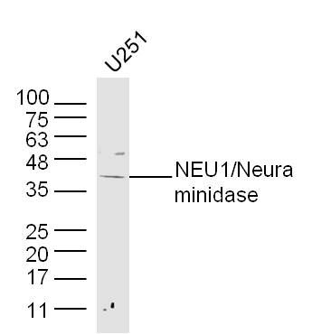
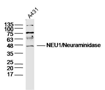
NEU1/Neuraminidase Polyclonal Antibody
Purified Rabbit Polyclonal Antibody (Pab)
- 产品详情
- 实验流程
Application
| WB, IHC-P, IHC-F, IF, ICC, E |
|---|---|
| Primary Accession | Q99519 |
| Reactivity | Rat, Bovine |
| Host | Rabbit |
| Clonality | Polyclonal |
| Calculated MW | 45467 Da |
| Physical State | Liquid |
| Immunogen | KLH conjugated synthetic peptide derived from human NEU1/Neuraminidase |
| Epitope Specificity | 151-250/415 |
| Isotype | IgG |
| Purity | affinity purified by Protein A |
| Buffer | 0.01M TBS (pH7.4) with 1% BSA, 0.02% Proclin300 and 50% Glycerol. |
| SUBCELLULAR LOCATION | Lysosome membrane. Lysosome lumen. Cell membrane. Cytoplasmic vesicle. Localized not only on the inner side of the lysosomal membrane and in the lysosomal lumen, but also on the plasma membrane and in intracellular vesicles. |
| SIMILARITY | Belongs to the glycosyl hydrolase 33 family. Contains 4 BNR repeats. |
| SUBUNIT | Interacts with cathepsin A (protective protein),beta-galactosidase and N-acetylgalactosamine-6-sulfate sulfatase in a multienzyme complex. |
| Post-translational modifications | N-glycosylated. Phosphorylation of tyrosine within the internalization signal results in inhibition of sialidase internalization and blockage on the plasma membrane. |
| DISEASE | Defects in NEU1 are the cause of sialidosis (SIALIDOSIS) [MIM:256550]. It is a lysosomal storage disease occurring as two types with various manifestations. Type 1 sialidosis (cherry red spot-myoclonus syndrome or normosomatic type) is late-onset and it is characterized by the formation of cherry red macular spots in childhood, progressive debilitating myoclonus, insiduous visual loss and rarely ataxia. The diagnosis can be confirmed by the screening of the urine for sialyloligosaccharides. Type 2 sialidosis (also known as dysmorphic type) occurs as several variants of increasing severity with earlier age of onset. It is characterized by the presence of abnormal somatic features including coarse facies and dysostosis multiplex, vertebral deformities, mental retardation, cherry-red spot/myoclonus, sialuria, cytoplasmic vacuolation of peripheral lymphocytes, bone marrow cells and conjunctival epithelial cells. |
| Important Note | This product as supplied is intended for research use only, not for use in human, therapeutic or diagnostic applications. |
| Background Descriptions | The protein encoded by this gene is a lysosomal enzyme that cleaves terminal sialic acid residues from substrates such as glycoproteins and glycolipids. In the lysosome, this enzyme is part of a heterotrimeric complex together with beta-galactosidase and cathepsin A (the latter is also referred to as 'protective protein'). Mutations in this gene can lead to sialidosis, a lysosomal storage disease that can be type 1 (cherry red spot-myoclonus syndrome or normosomatic type), which is late-onset, or type 2 (the dysmorphic type), which occurs at an earlier age with increased severity. [provided by RefSeq, Jul 2008] |
| Gene ID | 4758 |
|---|---|
| Other Names | Sialidase-1, 3.2.1.18, Acetylneuraminyl hydrolase, G9 sialidase, Lysosomal sialidase, N-acetyl-alpha-neuraminidase 1, NEU1, NANH |
| Target/Specificity | Highly expressed in pancreas, followed by skeletal muscle, kidney, placenta, heart, lung and liver. Weakly expressed in brain. |
| Dilution | WB=1:500-2000,IHC-P=1:100-500,IHC-F=1:100-500,ICC=1:100-500,IF=1:100-500,ELISA=1:5000-10000 |
| Format | 0.01M TBS(pH7.4) with 1% BSA, 0.09% (W/V) sodium azide and 50% Glyce |
| Storage | Store at -20 °C for one year. Avoid repeated freeze/thaw cycles. When reconstituted in sterile pH 7.4 0.01M PBS or diluent of antibody the antibody is stable for at least two weeks at 2-4 °C. |
| Name | NEU1 |
|---|---|
| Synonyms | NANH |
| Function | Catalyzes the removal of sialic acid (N-acetylneuraminic acid) moieties from glycoproteins and glycolipids. To be active, it is strictly dependent on its presence in the multienzyme complex. Appears to have a preference for alpha 2-3 and alpha 2-6 sialyl linkage. |
| Cellular Location | Lysosome membrane; Peripheral membrane protein; Lumenal side. Lysosome lumen. Cell membrane. Cytoplasmic vesicle Lysosome. Note=Localized not only on the inner side of the lysosomal membrane and in the lysosomal lumen, but also on the plasma membrane and in intracellular vesicles |
| Tissue Location | Highly expressed in pancreas, followed by skeletal muscle, kidney, placenta, heart, lung and liver. Weakly expressed in brain. |
Research Areas
For Research Use Only. Not For Use In Diagnostic Procedures.
Application Protocols
Provided below are standard protocols that you may find useful for product applications.
终于等到您。ABCEPTA(百远生物)抗体产品。
点击下方“我要评价 ”按钮提交您的反馈信息,您的反馈和评价是我们最宝贵的财富之一,
我们将在1-3个工作日内处理您的反馈信息。
如有疑问,联系:0512-88856768 tech-china@abcepta.com.
¥ 1,500.00
Cat# AP59051


