


 癌症的基本特征包括细胞增殖、血管生成、迁移、凋亡逃避机制和细胞永生等。找到癌症发生过程中这些通路的关键标记物和对应的抗体用于检测至关重要。
 为您推荐一个泛素化位点预测神器——泛素化分析工具,可以为您的蛋白的泛素化位点作出预测和评分。
 细胞自噬受体图形绘图工具为你的蛋白的细胞受体结合位点作出预测和评分,识别结合到自噬通路中的蛋白是非常重要的,便于让我们理解自噬在正常生理、病理过程中的作用,如发育、细胞分化、神经退化性疾病、压力条件下、感染和癌症。
CAF1p55/VPRBP Rabbit pAb
CAF1p55/VPRBP Rabbit pAb
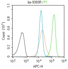
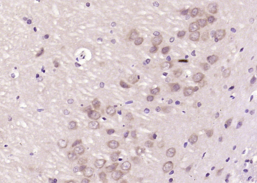
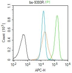
- 产品详情
- 实验流程
- 背景知识
Application 
| IHC-P, IHC-F, IF |
|---|---|
| Primary Accession | Q9Y4B6 |
| Reactivity | Rat, Human |
| Predicted | Mouse, Rabbit |
| Host | Rabbit |
| Clonality | Polyclonal |
| Calculated MW | 169007 Da |
| Physical State | Liquid |
| Immunogen | KLH conjugated synthetic peptide derived from human p55/DCAF1 |
| Epitope Specificity | 221-320/1507 |
| Isotype | IgG |
| Purity | affinity purified by Protein A |
| Buffer | 0.01M TBS (pH7.4) with 1% BSA, 0.02% Proclin300 and 50% Glycerol. |
| SUBCELLULAR LOCATION | Cytoplasm. Nucleus. |
| Important Note | This product as supplied is intended for research use only, not for use in human, therapeutic or diagnostic applications. |
| Background Descriptions | P55 is an extensively palmitoylated erythrocyte membrane protein, and a member of the MAGUK family. P55 also resists salt extraction, resulting in a high affinity for the plasma membrane. P55 contains a PDZ/DHR domain, a conserved SH-3 domain that appears to suppress tyrosine kinase activity of various oncoproteins, a 39-amino acid motif that binds to cytoskeletal protein 4.1R, and a guanylate kinase-like domain. Interaction with glycophorin C (GPC) and 4.1R suggests that p55 may play a role in the dynamic regulation in the erythrocyte membrane. In addition, p55 gene expression in vivo may be associated with a CpG island. P55 is constitutively expressed in K562 erythroleukemia cells during erythropoiesis and undergoes a 2-fold amplification after induction. |
| Gene ID | 9730 |
|---|---|
| Other Names | DDB1- and CUL4-associated factor 1 {ECO:0000312|HGNC:HGNC:30911}, HIV-1 Vpr-binding protein, VprBP, Serine/threonine-protein kinase VPRBP, 2.7.11.1, Vpr-interacting protein, DCAF1 (HGNC:30911), KIAA0800, RIP, VPRBP |
| Dilution | IHC-P=1:100-500,IHC-F=1:100-500,IF=1:100-500,Flow-Cyt=1ug/Test |
| Storage | Store at -20 °C for one year. Avoid repeated freeze/thaw cycles. When reconstituted in sterile pH 7.4 0.01M PBS or diluent of antibody the antibody is stable for at least two weeks at 2-4 °C. |
For Research Use Only. Not For Use In Diagnostic Procedures.
| Name | DCAF1 (HGNC:30911) |
|---|---|
| Synonyms | KIAA0800, RIP, VPRBP |
| Function | Acts both as a substrate recognition component of E3 ubiquitin-protein ligase complexes and as an atypical serine/threonine- protein kinase, playing key roles in various processes such as cell cycle, telomerase regulation and histone modification. Probable substrate-specific adapter of a DCX (DDB1-CUL4-X-box) E3 ubiquitin- protein ligase complex, named CUL4A-RBX1-DDB1-DCAF1/VPRBP complex, which mediates ubiquitination and proteasome-dependent degradation of proteins such as NF2 (PubMed:23063525). Involved in the turnover of methylated proteins: recognizes and binds methylated proteins via its chromo domain, leading to ubiquitination of target proteins by the RBX1-DDB1-DCAF1/VPRBP complex (PubMed:23063525). The CUL4A-RBX1-DDB1- DCAF1/VPRBP complex is also involved in B-cell development: DCAF1 is recruited by RAG1 to ubiquitinate proteins, leading to limit error- prone repair during V(D)J recombination (By similarity). Also part of the EDVP complex, an E3 ligase complex that mediates ubiquitination of proteins such as TERT, leading to TERT degradation and telomerase inhibition (PubMed:19287380, PubMed:23362280). The EDVP complex also mediates ubiquitination and degradation of CCP110 (PubMed:28242748, PubMed:34259627). Also acts as an atypical serine/threonine-protein kinase that specifically mediates phosphorylation of 'Thr-120' of histone H2A (H2AT120ph) in a nucleosomal context, thereby repressing transcription (PubMed:24140421). H2AT120ph is present in the regulatory region of many tumor suppresor genes, down-regulates their transcription and is present at high level in a number of tumors (PubMed:24140421). Involved in JNK-mediated apoptosis during cell competition process via its interaction with LLGL1 and LLGL2 (PubMed:20644714). By acting on TET dioxygenses, essential for oocyte maintenance at the primordial follicle stage, hence essential for female fertility (By similarity). |
| Cellular Location | Cytoplasm. Nucleus. Cytoplasm, cytoskeleton, microtubule organizing center, centrosome. Note=Associated with chromatin in a DDB1- independent and cell cycle-dependent manner: recruited to chromatin as DNA is being replicated and is released from chromatin before mitosis (By similarity). Homogeneous pancellular distribution is observed outside S-phase and a slight cytoplasmic-to-nuclear translocation from early to late S-phase (By similarity). Colocalizes with TET1 and PCNA at replicating heterochromatin during late S phase (By similarity) More concentrated in nuclei than in cytoplasm in germinal vesicle (GV) stage oocytes, zygotes and the 2-cell stage, but distributed in the cytoplasm at the MII-stage oocytes (By similarity). Localizes to centrosomes following interaction with CEP78 (PubMed:28242748, PubMed:34259627). {ECO:0000250|UniProtKB:Q80TR8, ECO:0000269|PubMed:28242748, ECO:0000269|PubMed:34259627} |
| Tissue Location | Ubiquitously expressed. |
Research Areas
Application Protocols
Provided below are standard protocols that you may find useful for product applications.
BACKGROUND
This product as supplied is intended for research use only, not for use in human, therapeutic or diagnostic applications.

终于等到您。ABCEPTA(百远生物)抗体产品。
点击下方“我要评价 ”按钮提交您的反馈信息,您的反馈和评价是我们最宝贵的财富之一,
我们将在1-3个工作日内处理您的反馈信息。
如有疑问,联系:0512-88856768 tech-china@abcepta.com.


