


 癌症的基本特征包括细胞增殖、血管生成、迁移、凋亡逃避机制和细胞永生等。找到癌症发生过程中这些通路的关键标记物和对应的抗体用于检测至关重要。
 为您推荐一个泛素化位点预测神器——泛素化分析工具,可以为您的蛋白的泛素化位点作出预测和评分。
 细胞自噬受体图形绘图工具为你的蛋白的细胞受体结合位点作出预测和评分,识别结合到自噬通路中的蛋白是非常重要的,便于让我们理解自噬在正常生理、病理过程中的作用,如发育、细胞分化、神经退化性疾病、压力条件下、感染和癌症。
Anti-ERK2 Antibody
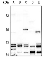
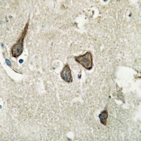
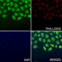
- 产品详情
- 实验流程
Application 
| WB, IHC, IF/IC |
|---|---|
| Primary Accession | P28482 |
| Reactivity | Human, Mouse, Rat, Zebrafish, Pig, Chicken, Bovine |
| Host | Rabbit |
| Clonality | Polyclonal |
| Calculated MW | 41390 Da |
| Gene ID | 5594 |
|---|---|
| Other Names | ERK2; PRKM1; PRKM2; Mitogen-activated protein kinase 1; MAP kinase 1; MAPK 1; ERT1; Extracellular signal-regulated kinase 2; ERK-2; MAP kinase isoform p42; p42-MAPK; Mitogen-activated protein kinase 2; MAP kinase 2; MAPK 2 |
| Target/Specificity | KLH-conjugated synthetic peptide encompassing a sequence within the center region of human ERK2. The exact sequence is proprietary. |
| Dilution | WB~~WB (1/500 - 1/1000) IHC~~1:100~500 IF/IC~~N/A |
| Format | Liquid in 0.42% Potassium phosphate, 0.87% Sodium chloride, pH 7.3, 30% glycerol, and 0.01% sodium azide. |
| Storage | Store at -20 °C.Stable for 12 months from date of receipt |
For Research Use Only. Not For Use In Diagnostic Procedures.
| Name | MAPK1 (HGNC:6871) |
|---|---|
| Synonyms | ERK2, PRKM1, PRKM2 |
| Function | Serine/threonine kinase which acts as an essential component of the MAP kinase signal transduction pathway. MAPK1/ERK2 and MAPK3/ERK1 are the 2 MAPKs which play an important role in the MAPK/ERK cascade. They participate also in a signaling cascade initiated by activated KIT and KITLG/SCF. Depending on the cellular context, the MAPK/ERK cascade mediates diverse biological functions such as cell growth, adhesion, survival and differentiation through the regulation of transcription, translation, cytoskeletal rearrangements. The MAPK/ERK cascade also plays a role in initiation and regulation of meiosis, mitosis, and postmitotic functions in differentiated cells by phosphorylating a number of transcription factors. About 160 substrates have already been discovered for ERKs. Many of these substrates are localized in the nucleus, and seem to participate in the regulation of transcription upon stimulation. However, other substrates are found in the cytosol as well as in other cellular organelles, and those are responsible for processes such as translation, mitosis and apoptosis. Moreover, the MAPK/ERK cascade is also involved in the regulation of the endosomal dynamics, including lysosome processing and endosome cycling through the perinuclear recycling compartment (PNRC); as well as in the fragmentation of the Golgi apparatus during mitosis. The substrates include transcription factors (such as ATF2, BCL6, ELK1, ERF, FOS, HSF4 or SPZ1), cytoskeletal elements (such as CANX, CTTN, GJA1, MAP2, MAPT, PXN, SORBS3 or STMN1), regulators of apoptosis (such as BAD, BTG2, CASP9, DAPK1, IER3, MCL1 or PPARG), regulators of translation (such as EIF4EBP1 and FXR1) and a variety of other signaling-related molecules (like ARHGEF2, DCC, FRS2, GLI1 or GRB10) (PubMed:35831023). Protein kinases (such as RAF1, RPS6KA1/RSK1, RPS6KA3/RSK2, RPS6KA2/RSK3, RPS6KA6/RSK4, SYK, MKNK1/MNK1, MKNK2/MNK2, RPS6KA5/MSK1, RPS6KA4/MSK2, MAPKAPK3 or MAPKAPK5) and phosphatases (such as DUSP1, DUSP4, DUSP6 or DUSP16) are other substrates which enable the propagation the MAPK/ERK signal to additional cytosolic and nuclear targets, thereby extending the specificity of the cascade. Mediates phosphorylation of TPR in response to EGF stimulation. May play a role in the spindle assembly checkpoint. Phosphorylates PML and promotes its interaction with PIN1, leading to PML degradation. Phosphorylates CDK2AP2 (By similarity). Phosphorylates phosphoglycerate kinase PGK1 under hypoxic conditions to promote its targeting to the mitochondrion and suppress the formation of acetyl-coenzyme A from pyruvate (PubMed:26942675). Phosphorylates GJA1 at 'Ser-279' and 'Ser- 282' resulting in an increase in GJA1 ubiquitination and ultimately lysosomal degradation (By similarity). Acts as a positive regulator of smoothened signaling by mediating phosphorylation of GLI1 in response to smoothened activation, promoting its dissociation from SUFU inhibitor and translocation to the nucleus (PubMed:35831023). |
| Cellular Location | Cytoplasm, cytoskeleton, spindle. Nucleus. Cytoplasm, cytoskeleton, microtubule organizing center, centrosome. Cytoplasm. Membrane, caveola {ECO:0000250|UniProtKB:P63086}. Cell junction, focal adhesion {ECO:0000250|UniProtKB:P63085}. Note=Associated with the spindle during prometaphase and metaphase (By similarity). PEA15-binding and phosphorylated DAPK1 promote its cytoplasmic retention. Phosphorylation at Ser- 246 and Ser-248 as well as autophosphorylation at Thr-190 promote nuclear localization. |
Research Areas
Application Protocols
Provided below are standard protocols that you may find useful for product applications.

终于等到您。ABCEPTA(百远生物)抗体产品。
点击下方“我要评价 ”按钮提交您的反馈信息,您的反馈和评价是我们最宝贵的财富之一,
我们将在1-3个工作日内处理您的反馈信息。
如有疑问,联系:0512-88856768 tech-china@abcepta.com.
¥ 1,250.00
Cat# AP60044


