


 癌症的基本特征包括细胞增殖、血管生成、迁移、凋亡逃避机制和细胞永生等。找到癌症发生过程中这些通路的关键标记物和对应的抗体用于检测至关重要。
 为您推荐一个泛素化位点预测神器——泛素化分析工具,可以为您的蛋白的泛素化位点作出预测和评分。
 细胞自噬受体图形绘图工具为你的蛋白的细胞受体结合位点作出预测和评分,识别结合到自噬通路中的蛋白是非常重要的,便于让我们理解自噬在正常生理、病理过程中的作用,如发育、细胞分化、神经退化性疾病、压力条件下、感染和癌症。
Anti-OPA1 Antibody
Rabbit polyclonal antibody to OPA1
- 产品详情
- 实验流程
- 背景知识
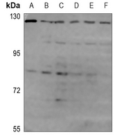
Application
| WB |
|---|---|
| Primary Accession | O60313 |
| Other Accession | P58281 |
| Reactivity | Human, Mouse, Rat |
| Host | Rabbit |
| Clonality | Polyclonal |
| Calculated MW | 111631 Da |
| Gene ID | 4976 |
|---|---|
| Other Names | KIAA0567; Dynamin-like 120 kDa protein mitochondrial; Optic atrophy protein 1 |
| Target/Specificity | KLH-conjugated synthetic peptide encompassing a sequence within the C-term region of human OPA1. The exact sequence is proprietary. |
| Dilution | WB~~WB (1/500 - 1/1000) |
| Format | Liquid in 0.42% Potassium phosphate, 0.87% Sodium chloride, pH 7.3, 30% glycerol, and 0.09% (W/V) sodium azide. |
| Storage | Store at -20 °C.Stable for 12 months from date of receipt |
| Name | OPA1 |
|---|---|
| Function | Dynamin-related GTPase that is essential for normal mitochondrial morphology by mediating fusion of the mitochondrial inner membranes, regulating cristae morphology and maintaining respiratory chain function (PubMed:16778770, PubMed:17709429, PubMed:20185555, PubMed:24616225, PubMed:28628083, PubMed:28746876, PubMed:31922487, PubMed:32228866, PubMed:32567732, PubMed:33130824, PubMed:33237841, PubMed:37612504, PubMed:37612506). Exists in two forms: the transmembrane, long form (Dynamin-like GTPase OPA1, long form; L-OPA1), which is tethered to the inner mitochondrial membrane, and the short soluble form (Dynamin-like GTPase OPA1, short form; S-OPA1), which results from proteolytic cleavage and localizes in the intermembrane space (PubMed:31922487, PubMed:32228866, PubMed:33237841, PubMed:37612504, PubMed:37612506). Both forms (L-OPA1 and S-OPA1) cooperate to catalyze the fusion of the mitochondrial inner membrane (PubMed:31922487, PubMed:37612504, PubMed:37612506). The equilibrium between L-OPA1 and S-OPA1 is essential: excess levels of S-OPA1, produced by cleavage by OMA1 following loss of mitochondrial membrane potential, lead to an impaired equilibrium between L-OPA1 and S-OPA1, inhibiting mitochondrial fusion (PubMed:20038677, PubMed:31922487). The balance between L-OPA1 and S-OPA1 also influences cristae shape and morphology (By similarity). Involved in remodeling cristae and the release of cytochrome c during apoptosis (By similarity). Proteolytic processing by PARL in response to intrinsic apoptotic signals may lead to disassembly of OPA1 oligomers and release of the caspase activator cytochrome C (CYCS) into the mitochondrial intermembrane space (By similarity). Acts as a regulator of T-helper Th17 cells, which are characterized by cells with fused mitochondria with tight cristae, by mediating mitochondrial membrane remodeling: OPA1 is required for interleukin-17 (IL-17) production (By similarity). Its role in mitochondrial morphology is required for mitochondrial genome maintenance (PubMed:18158317, PubMed:20974897). |
| Cellular Location | [Dynamin-like GTPase OPA1, long form]: Mitochondrion inner membrane; Single-pass membrane protein. Note=Detected at contact sites between endoplasmic reticulum and mitochondrion membranes. |
| Tissue Location | Highly expressed in retina (PubMed:11017079, PubMed:11017080, PubMed:11810270). Also expressed in brain, testis, heart and skeletal muscle (PubMed:11810270). Low levels of all isoforms expressed in a variety of tissues (PubMed:11810270) [Isoform 2]: Isoform 2 expressed in colon, liver, kidney, thyroid gland and leukocytes. |
Research Areas
For Research Use Only. Not For Use In Diagnostic Procedures.
Application Protocols
Provided below are standard protocols that you may find useful for product applications.
BACKGROUND
KLH-conjugated synthetic peptide encompassing a sequence within the C-term region of human OPA1. The exact sequence is proprietary.
终于等到您。ABCEPTA(百远生物)抗体产品。
点击下方“我要评价 ”按钮提交您的反馈信息,您的反馈和评价是我们最宝贵的财富之一,
我们将在1-3个工作日内处理您的反馈信息。
如有疑问,联系:0512-88856768 tech-china@abcepta.com.
¥ 1,500.00
Cat# AP60195


