


 癌症的基本特征包括细胞增殖、血管生成、迁移、凋亡逃避机制和细胞永生等。找到癌症发生过程中这些通路的关键标记物和对应的抗体用于检测至关重要。
 为您推荐一个泛素化位点预测神器——泛素化分析工具,可以为您的蛋白的泛素化位点作出预测和评分。
 细胞自噬受体图形绘图工具为你的蛋白的细胞受体结合位点作出预测和评分,识别结合到自噬通路中的蛋白是非常重要的,便于让我们理解自噬在正常生理、病理过程中的作用,如发育、细胞分化、神经退化性疾病、压力条件下、感染和癌症。
Anti-GNAT1 Antibody
Rabbit polyclonal antibody to GNAT1
- 产品详情
- 实验流程
- 背景知识
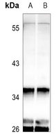
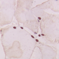
Application
| WB, IHC |
|---|---|
| Primary Accession | P11488 |
| Other Accession | P20612 |
| Reactivity | Human, Mouse, Rat, Bovine, Drosophila |
| Host | Rabbit |
| Clonality | Polyclonal |
| Calculated MW | 40041 Da |
| Gene ID | 2779 |
|---|---|
| Other Names | GNATR; Guanine nucleotide-binding protein G(t) subunit alpha-1; Transducin alpha-1 chain |
| Target/Specificity | KLH-conjugated synthetic peptide encompassing a sequence within the center region of human GNAT1. The exact sequence is proprietary. |
| Dilution | WB~~WB (1/500 - 1/1000), IHC (1/100 - 1/200) IHC~~WB (1/500 - 1/1000), IHC (1/100 - 1/200) |
| Format | Liquid in 0.42% Potassium phosphate, 0.87% Sodium chloride, pH 7.3, 30% glycerol, and 0.09% (W/V) sodium azide. |
| Storage | Store at -20 °C.Stable for 12 months from date of receipt |
| Name | GNAT1 |
|---|---|
| Synonyms | GNATR |
| Function | Functions as a signal transducer for the rod photoreceptor RHO. Required for normal RHO-mediated light perception by the retina (PubMed:22190596). Guanine nucleotide-binding proteins (G proteins) function as transducers downstream of G protein-coupled receptors (GPCRs), such as the photoreceptor RHO. The alpha chain contains the guanine nucleotide binding site and alternates between an active, GTP- bound state and an inactive, GDP-bound state. Activated RHO promotes GDP release and GTP binding. Signaling is mediated via downstream effector proteins, such as cGMP-phosphodiesterase (By similarity). |
| Cellular Location | Cell projection, cilium, photoreceptor outer segment {ECO:0000250|UniProtKB:P04695}. Membrane {ECO:0000250|UniProtKB:P04695}; Peripheral membrane protein {ECO:0000250|UniProtKB:P04695}. Photoreceptor inner segment {ECO:0000250|UniProtKB:P20612}. Note=Localizes mainly in the outer segment in the dark-adapted state, whereas is translocated to the inner part of the photoreceptors in the light-adapted state. During dark- adapted conditions, in the presence of UNC119 mislocalizes from the outer segment to the inner part of rod photoreceptors which leads to decreased photoreceptor damage caused by light {ECO:0000250|UniProtKB:P20612} |
| Tissue Location | Rod photoreceptor cells (PubMed:1614872). Predominantly expressed in the retina followed by the ciliary body, iris and retinal pigment epithelium (PubMed:22190596) |
Research Areas
For Research Use Only. Not For Use In Diagnostic Procedures.
Application Protocols
Provided below are standard protocols that you may find useful for product applications.
BACKGROUND
KLH-conjugated synthetic peptide encompassing a sequence within the center region of human GNAT1. The exact sequence is proprietary.
终于等到您。ABCEPTA(百远生物)抗体产品。
点击下方“我要评价 ”按钮提交您的反馈信息,您的反馈和评价是我们最宝贵的财富之一,
我们将在1-3个工作日内处理您的反馈信息。
如有疑问,联系:0512-88856768 tech-china@abcepta.com.


