


 癌症的基本特征包括细胞增殖、血管生成、迁移、凋亡逃避机制和细胞永生等。找到癌症发生过程中这些通路的关键标记物和对应的抗体用于检测至关重要。
 为您推荐一个泛素化位点预测神器——泛素化分析工具,可以为您的蛋白的泛素化位点作出预测和评分。
 细胞自噬受体图形绘图工具为你的蛋白的细胞受体结合位点作出预测和评分,识别结合到自噬通路中的蛋白是非常重要的,便于让我们理解自噬在正常生理、病理过程中的作用,如发育、细胞分化、神经退化性疾病、压力条件下、感染和癌症。
Anti-PHLDA1 Antibody
Rabbit polyclonal antibody to PHLDA1
- 产品详情
- 实验流程
- 背景知识
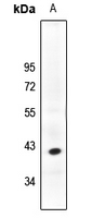
Application
| WB |
|---|---|
| Primary Accession | Q8WV24 |
| Other Accession | Q62392 |
| Reactivity | Human, Mouse, Rat, Bovine |
| Host | Rabbit |
| Clonality | Polyclonal |
| Calculated MW | 45016 Da |
| Gene ID | 22822 |
|---|---|
| Other Names | PHRIP; TDAG51; Pleckstrin homology-like domain family A member 1; Apoptosis-associated nuclear protein; Proline- and glutamine-rich protein; PQ-rich protein; PQR protein; Proline- and histidine-rich protein; T-cell death-associated gene 51 protein |
| Target/Specificity | KLH-conjugated synthetic peptide encompassing a sequence within the C-term region of human PHLDA1. The exact sequence is proprietary. |
| Dilution | WB~~WB (1/500 - 1/1000) |
| Format | Liquid in 0.42% Potassium phosphate, 0.87% Sodium chloride, pH 7.3, 30% glycerol, and 0.09% (W/V) sodium azide. |
| Storage | Store at -20 °C.Stable for 12 months from date of receipt |
| Name | PHLDA1 |
|---|---|
| Synonyms | PHRIP, TDAG51 |
| Function | Seems to be involved in regulation of apoptosis. May be involved in detachment-mediated programmed cell death. May mediate apoptosis during neuronal development. May be involved in regulation of anti-apoptotic effects of IGF1. May be involved in translational regulation. |
| Cellular Location | Cytoplasm. Cytoplasmic vesicle. Nucleus, nucleolus. Note=Colocalizes with intracellular vesicles. |
| Tissue Location | Widely expressed with highest levels in pancreas. Strongly expressed by benign melanocytic nevi, and progressively reduced expressed in primary and metastatic melanomas (at protein level). |
Research Areas
For Research Use Only. Not For Use In Diagnostic Procedures.
Application Protocols
Provided below are standard protocols that you may find useful for product applications.
BACKGROUND
KLH-conjugated synthetic peptide encompassing a sequence within the C-term region of human PHLDA1. The exact sequence is proprietary.
终于等到您。ABCEPTA(百远生物)抗体产品。
点击下方“我要评价 ”按钮提交您的反馈信息,您的反馈和评价是我们最宝贵的财富之一,
我们将在1-3个工作日内处理您的反馈信息。
如有疑问,联系:0512-88856768 tech-china@abcepta.com.
¥ 1,500.00
Cat# AP61203


