


 癌症的基本特征包括细胞增殖、血管生成、迁移、凋亡逃避机制和细胞永生等。找到癌症发生过程中这些通路的关键标记物和对应的抗体用于检测至关重要。
 为您推荐一个泛素化位点预测神器——泛素化分析工具,可以为您的蛋白的泛素化位点作出预测和评分。
 细胞自噬受体图形绘图工具为你的蛋白的细胞受体结合位点作出预测和评分,识别结合到自噬通路中的蛋白是非常重要的,便于让我们理解自噬在正常生理、病理过程中的作用,如发育、细胞分化、神经退化性疾病、压力条件下、感染和癌症。
Anti-CD4 (pS433) Antibody
Rabbit polyclonal antibody to CD4 (pS433)
- 产品详情
- 实验流程
- 背景知识
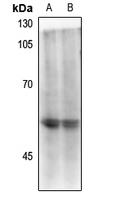
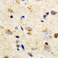
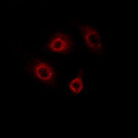
Application
| WB, IHC, IF |
|---|---|
| Primary Accession | P01730 |
| Other Accession | P06332 |
| Reactivity | Human, Mouse, Rat, Monkey, Drosophila |
| Host | Rabbit |
| Clonality | Polyclonal |
| Calculated MW | 51111 Da |
| Gene ID | 920 |
|---|---|
| Other Names | T-cell surface glycoprotein CD4; T-cell surface antigen T4/Leu-3; CD4 |
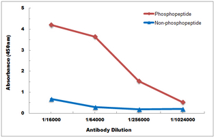
| Target/Specificity | KLH-conjugated synthetic peptide encompassing a sequence within the C-term region of human CD4 with a site at pS433. The exact sequence is proprietary. |
| Dilution | WB~~WB (1/500 - 1/1000), IH (1/50 - 1/200), IF/IC (1/50 - 1/200) IHC~~1:100~500 IF~~WB (1/500 - 1/1000), IH (1/50 - 1/200), IF/IC (1/50 - 1/200) |
| Format | Liquid in 0.42% Potassium phosphate, 0.87% Sodium chloride, pH 7.3, 30% glycerol, and 0.09% (W/V) sodium azide. |
| Storage | Store at -20 °C.Stable for 12 months from date of receipt |
| Name | CD4 |
|---|---|
| Function | Integral membrane glycoprotein that plays an essential role in the immune response and serves multiple functions in responses against both external and internal offenses. In T-cells, functions primarily as a coreceptor for MHC class II molecule:peptide complex. The antigens presented by class II peptides are derived from extracellular proteins while class I peptides are derived from cytosolic proteins. Interacts simultaneously with the T-cell receptor (TCR) and the MHC class II presented by antigen presenting cells (APCs). In turn, recruits the Src kinase LCK to the vicinity of the TCR-CD3 complex. LCK then initiates different intracellular signaling pathways by phosphorylating various substrates ultimately leading to lymphokine production, motility, adhesion and activation of T-helper cells. In other cells such as macrophages or NK cells, plays a role in differentiation/activation, cytokine expression and cell migration in a TCR/LCK-independent pathway. Participates in the development of T- helper cells in the thymus and triggers the differentiation of monocytes into functional mature macrophages. |
| Cellular Location | Cell membrane; Single-pass type I membrane protein. Note=Localizes to lipid rafts (PubMed:12517957, PubMed:9168119). Removed from plasma membrane by HIV- 1 Nef protein that increases clathrin-dependent endocytosis of this antigen to target it to lysosomal degradation. Cell surface expression is also down-modulated by HIV-1 Envelope polyprotein gp160 that interacts with, and sequesters CD4 in the endoplasmic reticulum |
| Tissue Location | Highly expressed in T-helper cells. The presence of CD4 is a hallmark of T-helper cells which are specialized in the activation and growth of cytotoxic T-cells, regulation of B cells, or activation of phagocytes. CD4 is also present in other immune cells such as macrophages, dendritic cells or NK cells |
Research Areas
For Research Use Only. Not For Use In Diagnostic Procedures.
Application Protocols
Provided below are standard protocols that you may find useful for product applications.
BACKGROUND
KLH-conjugated synthetic peptide encompassing a sequence within the C-term region of human CD4 with a site at pS433. The exact sequence is proprietary.
终于等到您。ABCEPTA(百远生物)抗体产品。
点击下方“我要评价 ”按钮提交您的反馈信息,您的反馈和评价是我们最宝贵的财富之一,
我们将在1-3个工作日内处理您的反馈信息。
如有疑问,联系:0512-88856768 tech-china@abcepta.com.
¥ 1,500.00
Cat# AP61448


