


 癌症的基本特征包括细胞增殖、血管生成、迁移、凋亡逃避机制和细胞永生等。找到癌症发生过程中这些通路的关键标记物和对应的抗体用于检测至关重要。
 为您推荐一个泛素化位点预测神器——泛素化分析工具,可以为您的蛋白的泛素化位点作出预测和评分。
 细胞自噬受体图形绘图工具为你的蛋白的细胞受体结合位点作出预测和评分,识别结合到自噬通路中的蛋白是非常重要的,便于让我们理解自噬在正常生理、病理过程中的作用,如发育、细胞分化、神经退化性疾病、压力条件下、感染和癌症。
OAS1 Antibody (C-term)
Purified Rabbit Polyclonal Antibody (Pab)
- 产品详情
- 文献引用 : 7
- 实验流程
- 背景知识
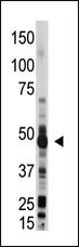
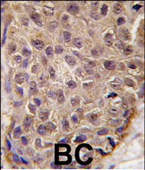
Application
| WB, IHC-P, E |
|---|---|
| Primary Accession | P00973 |
| Reactivity | Human, Mouse |
| Host | Rabbit |
| Clonality | Polyclonal |
| Isotype | Rabbit IgG |
| Calculated MW | 46029 Da |
| Antigen Region | 302-330 aa |
| Gene ID | 4938 |
|---|---|
| Other Names | 2'-5'-oligoadenylate synthase 1, (2-5')oligo(A) synthase 1, 2-5A synthase 1, E18/E16, p46/p42 OAS, OAS1, OIAS |
| Target/Specificity | This OAS1 antibody is generated from rabbits immunized with a KLH conjugated synthetic peptide between 302-330 amino acids from the C-terminal region of human OAS1. |
| Dilution | WB~~1:1000 IHC-P~~1:100~500 E~~Use at an assay dependent concentration. |
| Format | Purified polyclonal antibody supplied in PBS with 0.09% (W/V) sodium azide. This antibody is prepared by Saturated Ammonium Sulfate (SAS) precipitation followed by dialysis against PBS. |
| Storage | Maintain refrigerated at 2-8°C for up to 2 weeks. For long term storage store at -20°C in small aliquots to prevent freeze-thaw cycles. |
| Precautions | OAS1 Antibody (C-term) is for research use only and not for use in diagnostic or therapeutic procedures. |
| Name | OAS1 |
|---|---|
| Synonyms | OIAS |
| Function | Interferon-induced, dsRNA-activated antiviral enzyme which plays a critical role in cellular innate antiviral response (PubMed:34581622). In addition, it may also play a role in other cellular processes such as apoptosis, cell growth, differentiation and gene regulation. Synthesizes higher oligomers of 2'-5'-oligoadenylates (2-5A) from ATP which then bind to the inactive monomeric form of ribonuclease L (RNase L) leading to its dimerization and subsequent activation. Activation of RNase L leads to degradation of cellular as well as viral RNA, resulting in the inhibition of protein synthesis, thus terminating viral replication (PubMed:34145065, PubMed:34581622, PubMed:40010341). Involved in intercellular immune signaling that limits local spread of RNA virus infection and protects against tumorigenesis (PubMed:40010341). Can generate high levels of 2',5'- oligoadenylates in transformed cells, targeting them to innate and adaptive immunesurveillance mechanisms (PubMed:40010341). Can mediate the antiviral effect via the classical RNase L-dependent pathway or an alternative antiviral pathway independent of RNase L. The secreted form displays antiviral effect against vesicular stomatitis virus (VSV), herpes simplex virus type 2 (HSV-2), and encephalomyocarditis virus (EMCV) and stimulates the alternative antiviral pathway independent of RNase L. |
| Cellular Location | Cytoplasm. Mitochondrion. Nucleus. Microsome Endoplasmic reticulum. Secreted {ECO:0000250|UniProtKB:Q29599}. Note=Associated with different subcellular fractions such as mitochondrial, nuclear, and rough/smooth microsomal fractions. [Isoform p42]: Note=(Microbial infection) In SARS coronavirus-2/SARS-CoV-2 infected cells, since its not prenylated, is diffusely localized and unable to initiate a detectable block to SARS- CoV-2 replication. |
| Tissue Location | Expressed in lungs.. |
For Research Use Only. Not For Use In Diagnostic Procedures.

Provided below are standard protocols that you may find useful for product applications.
BACKGROUND
OAS1 is an interferon inducible protein that may play a role in mediating resistance to virus infection, control of cell growth, differentiation, and apoptosis. It binds double-stranded RNA and polymerizes ATP into PPP(A2'P5'A)N oligomers, which activate the latent RNase L that, when activated, cleaves single-stranded RNAs. This protein is associated with different subcellular fractions such as mitochondrial, nuclear, and rough/smooth microsomal fractions.
REFERENCES
Strausberg, R.L., et al., Proc. Natl. Acad. Sci. U.S.A. 99(26):16899-16903 (2002).
Sarkar, S.N., et al., J. Biol. Chem. 274(36):25535-25542 (1999).
Ghosh, A., et al., J. Biol. Chem. 272(52):33220-33226 (1997).
Ghosh, S.K., et al., J. Biol. Chem. 266(23):15293-15299 (1991).
Rutherford, M.N., et al., EMBO J. 7(3):751-759 (1988).
终于等到您。ABCEPTA(百远生物)抗体产品。
点击下方“我要评价 ”按钮提交您的反馈信息,您的反馈和评价是我们最宝贵的财富之一,
我们将在1-3个工作日内处理您的反馈信息。
如有疑问,联系:0512-88856768 tech-china@abcepta.com.


