


 癌症的基本特征包括细胞增殖、血管生成、迁移、凋亡逃避机制和细胞永生等。找到癌症发生过程中这些通路的关键标记物和对应的抗体用于检测至关重要。
 为您推荐一个泛素化位点预测神器——泛素化分析工具,可以为您的蛋白的泛素化位点作出预测和评分。
 细胞自噬受体图形绘图工具为你的蛋白的细胞受体结合位点作出预测和评分,识别结合到自噬通路中的蛋白是非常重要的,便于让我们理解自噬在正常生理、病理过程中的作用,如发育、细胞分化、神经退化性疾病、压力条件下、感染和癌症。
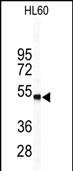
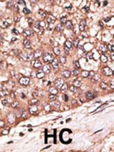
WISP1 Antibody (Center)
Purified Rabbit Polyclonal Antibody (Pab)
- 产品详情
- 文献引用 : 1
- 实验流程
- 背景知识
Application
| WB, IHC-P, E |
|---|---|
| Primary Accession | O95388 |
| Reactivity | Human |
| Host | Rabbit |
| Clonality | Polyclonal |
| Isotype | Rabbit IgG |
| Calculated MW | 40331 Da |
| Antigen Region | 171-200 aa |
| Gene ID | 8840 |
|---|---|
| Other Names | WNT1-inducible-signaling pathway protein 1, WISP-1, CCN family member 4, Wnt-1-induced secreted protein, WISP1, CCN4 |
| Target/Specificity | This WISP1 antibody is generated from rabbits immunized with a KLH conjugated synthetic peptide between 171-200 amino acids from the Central region of human WISP1. |
| Dilution | WB~~1:1000 IHC-P~~1:100~500 E~~Use at an assay dependent concentration. |
| Format | Purified polyclonal antibody supplied in PBS with 0.09% (W/V) sodium azide. This antibody is prepared by Saturated Ammonium Sulfate (SAS) precipitation followed by dialysis against PBS. |
| Storage | Maintain refrigerated at 2-8°C for up to 2 weeks. For long term storage store at -20°C in small aliquots to prevent freeze-thaw cycles. |
| Precautions | WISP1 Antibody (Center) is for research use only and not for use in diagnostic or therapeutic procedures. |
| Name | CCN4 (HGNC:12769) |
|---|---|
| Synonyms | WISP1 |
| Function | Downstream regulator in the Wnt/Frizzled-signaling pathway. Associated with cell survival. Attenuates p53-mediated apoptosis in response to DNA damage through activation of AKT kinase. Up-regulates the anti-apoptotic Bcl-X(L) protein. Adheres to skin and melanoma fibroblasts. In vitro binding to skin fibroblasts occurs through the proteoglycans, decorin and biglycan. |
| Cellular Location | Secreted. |
| Tissue Location | Expressed in heart, kidney, lung, pancreas, placenta, ovary, small intestine and spleen. Isoform 2 is expressed predominantly in scirrhous gastric carcinoma and, weakly in placenta Overexpression is associated with several cancers including breast cancer and colon tumors. Isoform 2 is overexpressed in scirrhous gastric carcinoma |
For Research Use Only. Not For Use In Diagnostic Procedures.

Provided below are standard protocols that you may find useful for product applications.
BACKGROUND
Wisp1 is a member of the WNT1 inducible signaling pathway (WISP) protein subfamily, which belongs to the connective tissue growth factor (CTGF) family. WNT1 is a member of a family of cysteine-rich, glycosylated signaling proteins that mediate diverse developmental processes. The CTGF family members are characterized by four conserved cysteine-rich domains: insulin-like growth factor-binding domain, von Willebrand factor type C module, thrombospondin domain and C-terminal cystine knot-like domain. Wisp1 may be downstream in the WNT1 signaling pathway that is relevant to malignant transformation. It is expressed at a high level in fibroblast cells, and overexpressed in colon tumors. The encoded protein binds to decorin and biglycan, two members of a family of small leucine-rich proteoglycans present in the extracellular matrix of connective tissue, and possibly prevents the inhibitory activity of decorin and biglycan in tumor cell proliferation. It also attenuates p53-mediated apoptosis in response to DNA damage through activation of the Akt kinase. It is 83% identical to the mouse protein at the amino acid level.
REFERENCES
Hocevar, B.A., et al., EMBO J. 22(12):3084-3094 (2003).
Tanaka, S., et al., Hepatology 37(5):1122-1129 (2003).
Soon, L.L., et al., J. Biol. Chem. 278(13):11465-11470 (2003).
Su, F., et al., Genes Dev. 16(1):46-57 (2002).
Xie, D., et al., Cancer Res. 61(24):8917-8923 (2001).
终于等到您。ABCEPTA(百远生物)抗体产品。
点击下方“我要评价 ”按钮提交您的反馈信息,您的反馈和评价是我们最宝贵的财富之一,
我们将在1-3个工作日内处理您的反馈信息。
如有疑问,联系:0512-88856768 tech-china@abcepta.com.


