


 癌症的基本特征包括细胞增殖、血管生成、迁移、凋亡逃避机制和细胞永生等。找到癌症发生过程中这些通路的关键标记物和对应的抗体用于检测至关重要。
 为您推荐一个泛素化位点预测神器——泛素化分析工具,可以为您的蛋白的泛素化位点作出预测和评分。
 细胞自噬受体图形绘图工具为你的蛋白的细胞受体结合位点作出预测和评分,识别结合到自噬通路中的蛋白是非常重要的,便于让我们理解自噬在正常生理、病理过程中的作用,如发育、细胞分化、神经退化性疾病、压力条件下、感染和癌症。
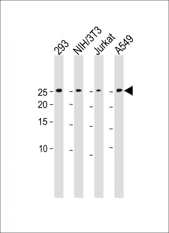
DDIT4 Antibody (C-term)
Purified Rabbit Polyclonal Antibody (Pab)

- 产品详情
- 实验流程
- 背景知识
Application
| WB, IHC-P, FC, E |
|---|---|
| Primary Accession | Q9NX09 |
| Other Accession | Q8VHZ9, Q9D3F7, Q08E62 |
| Reactivity | Human |
| Predicted | Mouse, Rat, Bovine |
| Host | Rabbit |
| Clonality | Polyclonal |
| Isotype | Rabbit IgG |
| Calculated MW | 25371 Da |
| Antigen Region | 198-227 aa |
| Gene ID | 54541 |
|---|---|
| Other Names | DNA damage-inducible transcript 4 protein, HIF-1 responsive protein RTP801, Protein regulated in development and DNA damage response 1, REDD-1, DDIT4, REDD1, RTP801 |
| Target/Specificity | This DDIT4 antibody is generated from rabbits immunized with a KLH conjugated synthetic peptide between 198-227 amino acids from the C-terminal region of human DDIT4. |
| Dilution | WB~~1:1000 IHC-P~~1:100~500 FC~~1:10~50 E~~Use at an assay dependent concentration. |
| Format | Purified polyclonal antibody supplied in PBS with 0.05% (V/V) Proclin 300. This antibody is prepared by Saturated Ammonium Sulfate (SAS) precipitation followed by dialysis against PBS. |
| Storage | Maintain refrigerated at 2-8°C for up to 2 weeks. For long term storage store at -20°C in small aliquots to prevent freeze-thaw cycles. |
| Precautions | DDIT4 Antibody (C-term) is for research use only and not for use in diagnostic or therapeutic procedures. |
| Name | DDIT4 |
|---|---|
| Synonyms | REDD1, RTP801 |
| Function | Regulates cell growth, proliferation and survival via inhibition of the activity of the mammalian target of rapamycin complex 1 (mTORC1). Inhibition of mTORC1 is mediated by a pathway that involves DDIT4/REDD1, AKT1, the TSC1-TSC2 complex and the GTPase RHEB. Plays an important role in responses to cellular energy levels and cellular stress, including responses to hypoxia and DNA damage. Regulates p53/TP53-mediated apoptosis in response to DNA damage via its effect on mTORC1 activity. Its role in the response to hypoxia depends on the cell type; it mediates mTORC1 inhibition in fibroblasts and thymocytes, but not in hepatocytes (By similarity). Required for mTORC1-mediated defense against viral protein synthesis and virus replication (By similarity). Inhibits neuronal differentiation and neurite outgrowth mediated by NGF via its effect on mTORC1 activity. Required for normal neuron migration during embryonic brain development. Plays a role in neuronal cell death. |
| Cellular Location | Mitochondrion. Cytoplasm, cytosol |
| Tissue Location | Broadly expressed, with lowest levels in brain, skeletal muscle and intestine. Up-regulated in substantia nigra neurons from Parkinson disease patients (at protein level) |
For Research Use Only. Not For Use In Diagnostic Procedures.
Provided below are standard protocols that you may find useful for product applications.
BACKGROUND
REDD1 is a novel transcriptional target of p53 induced following DNA damage. During embryogenesis, REDD1 expression mirrors the tissue-specific pattern of the p53 family member p63, and TP63 null embryos show virtually no expression of REDD1, which is restored in mouse embryo fibroblasts following p63 expression. In differentiating primary keratinocytes, TP63 and REDD1 expression are coordinately downregulated, and ectopic expression of either gene inhibits in vitro differentiation. REDD1 appears to function in the regulation of reactive oxygen species (ROS); TP63 null fibroblasts have decreased ROS levels and reduced sensitivity to oxidative stress, which are both increased following ectopic expression of either TP63 or REDD1. Thus, REDD1 encodes a shared transcriptional target that implicates ROS in the p53-dependent DNA damage response and in p63-mediated regulation of epithelial differentiation.
REFERENCES
Ellisen L.W., Mol. Cell 10:995-1005(2002).
Shoshani T.,Mol. Cell. Biol. 22:2283-2293(2002).
终于等到您。ABCEPTA(百远生物)抗体产品。
点击下方“我要评价 ”按钮提交您的反馈信息,您的反馈和评价是我们最宝贵的财富之一,
我们将在1-3个工作日内处理您的反馈信息。
如有疑问,联系:0512-88856768 tech-china@abcepta.com.


